
Presentation
Chest pain.
Patient Data
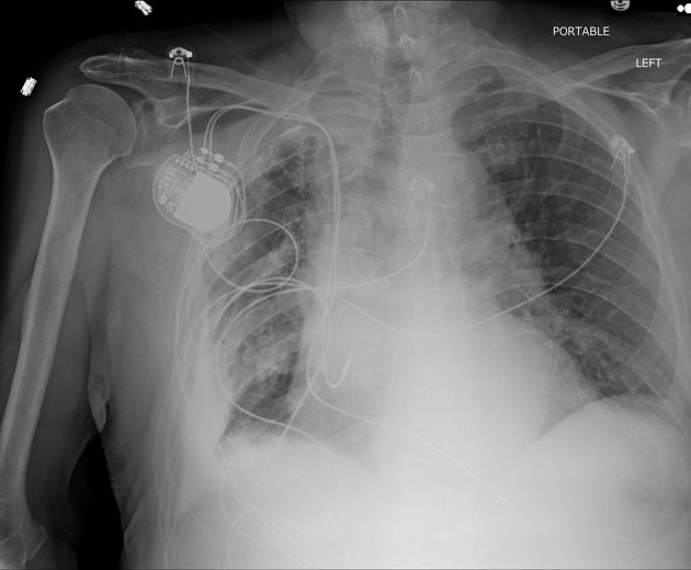
Left costophrenic angle is not included in the study.
Right-sided dual lead permanent pacemaker.
Volume loss of the right thorax with mild mediastinal shift to the right. Diffuse right pleural thickening, with calcific plaquing particularly at the right costophrenic angle. The visualized left lung is clear. No pneumothorax, vascular congestion, or pleural effusion. The cardiac silhouette is slightly enlarged. Aortic arch calcification is present.
Case Discussion
Findings are suggestive of a chronic right fibrothorax. The history of empyema, tuberculosis, hemothorax, rheumatoid, or asbestosis can help clench the diagnosis. Our patient had a history of right-sided hemothorax secondary to trauma.
Fibrothorax is a time-dependent process. It can be prevented if the pleural disease is treated in a timely manner. Depending on the cause, insertion of a chest tube can remove an effusion or hemothorax, preventing the development of a fibrothorax.
Sometimes a decortication procedure is required once the onset of fibrosis begins. This will allow the lung to reexpand.


 Unable to process the form. Check for errors and try again.