
Presentation
Severe left hip pain and limitation of movement after mild trauma
Patient Data
Age: 25 years
Gender: Male
From the case:
Fibrous dysplasia and pathological fracture


Download
Info
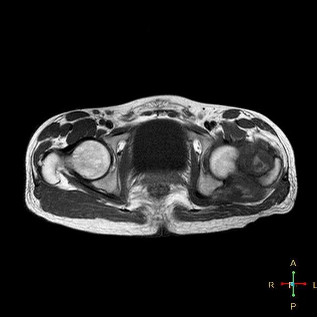
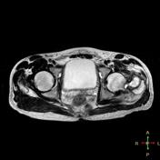
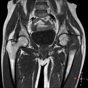
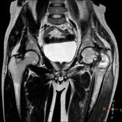
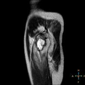
Intertrochanteric fracture of the left femur owing to presence of an oval shaped well circumscribed intramedullary lesion that displays low signal in T1 and bright signal in T2 and STIR with hypointense sclerotic rim. Marrow edema of the left femoral neck and contusion of adjacent muscles are noted. Fibrous dysplasia was found on biopsy.
Case Discussion
Pathological fractures may occur secondary to systemic disease affecting the bones and lead to demineralization e.g. osteoporosis and rickets or due to bone lesion e.g. simple bone cyst and fibrous dysplasia. When the lesion exceeds 75% of bone circumference it becomes liable to fracture.


 Unable to process the form. Check for errors and try again.