
Presentation
Referred for imaging. Patient with known left fibula hemimelia.
Patient Data
Age: 3.5 years
Gender: Female
From the case:
Fibular hemimelia
Download
Info
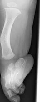
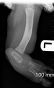
The left tibia is foreshortened, thickened and bowed anteromedially. The left fibula is absent. There is evidence of talipes equinovalgus deformity.
The lateral rays and phalanges (4th and 5th digits) are also absent.
Findings are consistent with type II fibular hemimelia.
Case Discussion
Although rare in occurrence, it is the most common congenital absence of long bones of the extremities.
Fibular hemimelia classification is that of Achterman and Kalamachi et al, which divides the condition into two types:
type I: minimal hypoplasia of the fibula
type II: complete absence of the fibula


 Unable to process the form. Check for errors and try again.