
Presentation
Abnormal LFT. Normal body habitus. Six years of prior oral contraceptive pill usage from 16 to 22 years.
Patient Data

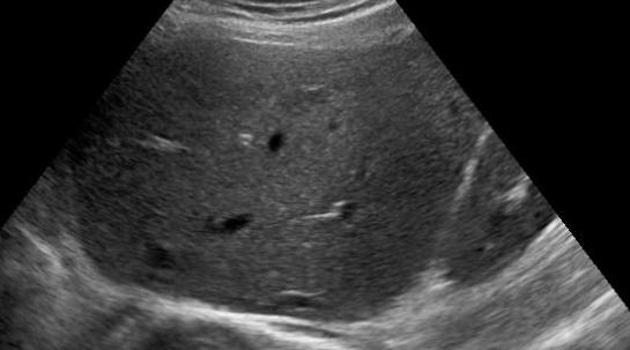
The liver appeared normal under the usual abdominal ultrasound settings. The gallbladder and bile ducts also appeared normal.
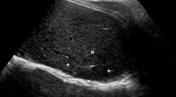
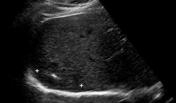
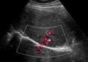
Liver with low dynamic range: an amorphous, hypoechoic region was visualized within segment VII after the dynamic range was lowered and the liver rescanned. There appeared to be an echogenic central scar. Blood flow appears to have a stellate distribution.
Case Discussion
Focal nodular hyperplasia is usually a very subtle finding on ultrasound unless there is a background of steatosis.
In this case, with an abnormal LFT and an otherwise normal appearing liver, what is being missed? I asked the patient about long-term OCP usage, dropped the dynamic range and had another look.
A hypoechoic area with a central scar and stellate blood distribution pattern was seen in segment VII. These findings point to focal nodular hyperplasia, which was confirmed with a CT scan.


 Unable to process the form. Check for errors and try again.