
Presentation
History of thorn injury to left middle finger palmer aspect about 2 weeks back. Complaining of finger edema along palmer side.
Patient Data
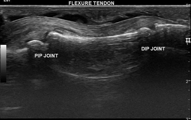
There is a linear echogenic focus (2.4 mm) in the subcutaneous plane of the mid-part of the proximal phalanx. It is ulnar to midline. There is surrounding tiny anechoic fluid rim. It is a foreign body - thorn fragment.
Local hypervascularity is present. There is effusion in flexor tendon sheath suggestive of tenosynovitis. Flexor tendons show normal echopattern. There is no joint effusion.
Case Discussion
A young male had thorn injury to the palmar aspect of the left middle finger. The patient presented with finger swelling. The patient was able to palpate a tiny nodule in the finger which turned out to be a thorn fragment in the subcutaneous plane. It was associated with flexor tendon tenosynovitis.
Surgical exploration revealed a thorn fragment at the expected location. Thorn removal and synovectomy were done. There was no foreign body in the tendon sheath. When a thorn pierces the tendon sheath, dirt enters into the sheath which causes synovitis. Flexure tenosynovitis of a finger is always 'bumpy' due to the presence of pulleys.


 Unable to process the form. Check for errors and try again.