
Presentation
Inappropriate behavior and difficulty at work, resulting in job loss.
Patient Data
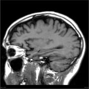

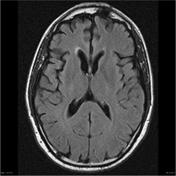
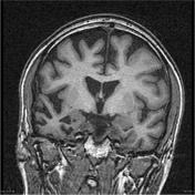
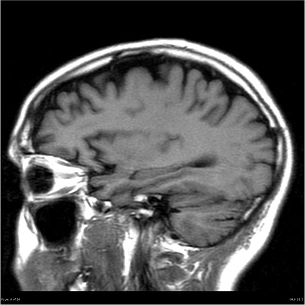
MRI demonstrates marked frontal and temporal atrophy, more marked on the right. There is also reduction in the caudate head size, again more pronounced on the right. Hippocampal volumes appear preserved.
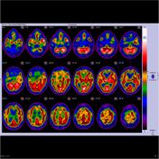
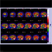
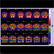
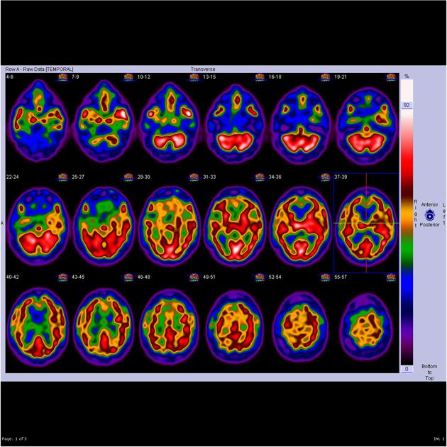
Studies were performed following sedation administered in the ward for difficult behavior.
SPECT studies performed following infusion of technetium HMPAO with mental stimulation and these of low statistical quality but appear to show mildly but extensively reduced perfusion to the frontal lobes, particularly along the mesial surfaces. Elsewhere perfusion appears normal, in particular, the posterior cingulate and precuneus hot zones are detected hence making early Alzheimer's disease unlikely. There is also hypoperfusion of the caudate heads bilaterally which can be seen Huntington's chorea, however, perfusion of the other basal ganglia, brainstem and cerebellum are normal.
Impression: Overall impression would favor a frontal impairment or dementia as the cause of the patient's behavior.
Case Discussion
Features of frontal and temporal atrophy, asymmetric, with caudate head volume loss are consistent with the clinical impression of a frontotemporal dementia.


 Unable to process the form. Check for errors and try again.