
Presentation
Breastfeeding patient with a a palpable mass in her breast
Patient Data

At initial presentation, the lesion is moderately heterogeneous on ultrasound. There is no skin thickening.
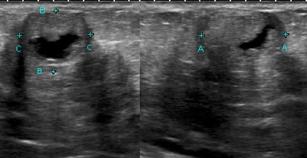
Two weeks later, the lesion shows some low-level echoes suggesting fluid in the lesion. At this stage, the differential diagnosis should include an abscess. No pain and no inflammation clinically.

The patient returns after 3 months for a follow-up study. The lesion is smaller and more echogenic. Aspiration showed inspissated white fluid.
Case Discussion
Galactoceles are lesions that have variable appearances on ultrasound. The differential diagnosis is malignancy, fibroadenoma, and abscess.
Careful follow up clinically and with imaging is essential not to miss potentially sinister lesions in the lactating breast. Be careful of simply assuming that lesions in the lactating breast are simply galactoceles. This is not true.


 Unable to process the form. Check for errors and try again.