
Presentation
Had cholecystectomy 2 years ago for gallbladder polyp with no follow-up.
Patient Data


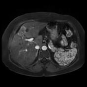
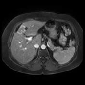
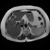
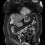
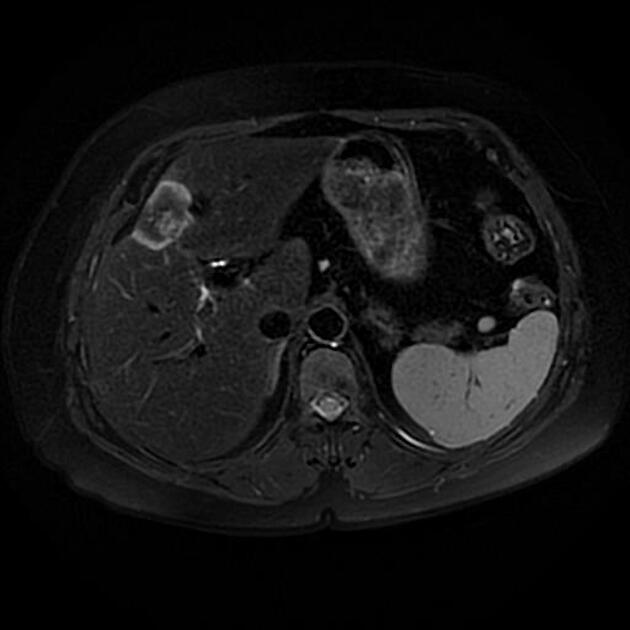
Dysmorphic liver, diffusely hyperintense on T1 IP, attenuated on T1 OP in keeping with diffuse hepatic steatosis.
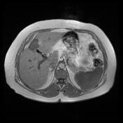
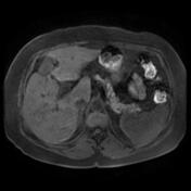
Hepatic mass centered on the gallbladder fossa with involvement of the segments (4 and 5). It elicits a low signal on T1 and T2 centrally and a high signal peripherally with heterogeneous enhancement on postcontrast sequences with hepatic capsular retraction. Small abdominal lymphadenopathy mainly of peripheral location (short axis ~ 8 mm).
Case Discussion
MRI features of a hepatic mass centered on the gallbladder fossa with involvement of the segments 4 and 5 of the liver, pathologically proven as a recurrence in a patient operated 2 years prior for a malignant gallbladder polyp (adenocarcinoma).


 Unable to process the form. Check for errors and try again.