
Presentation
On and off right upper quadrant pain radiating to the right shoulder for 5 years.
Patient Data

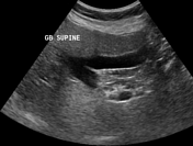
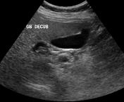
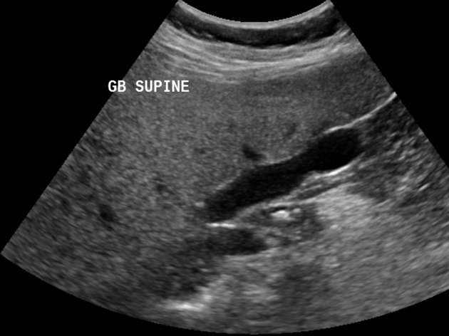
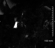
No gallstones are seen. No abnormal gallbladder wall thickening or pericholecystic fluid is seen as well.


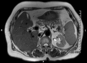
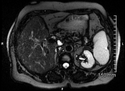
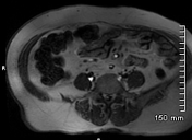
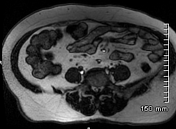
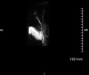
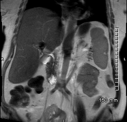
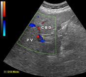
Enlarged liver (19 cm) with mild fatty infiltration. No focal hepatic lesion or biliary dilatation is seen. Fatty infiltration of the pancreas. Normal gallbladder with two septae; however, no gallstones or abnormal gall bladder wall thickening is noted. Normal caliber CBD and pancreatic duct. Ectopic right kidney seen in the pelvis. Small simple cyst at the upper pole of left kidney. A note is also made of left renal vein coursing obliquely and inferiorly to join the left common iliac vein (type IV renal vein).
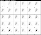
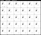
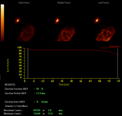

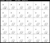

Radiopharmaceutical: 194 MBqs of Tc-99m BrIDA (mebrofenin trimethylbromo-iminodiaceticacid) injected IV as per protocol. Post-injection, immediate dynamic images of the liver and the gallbladder were acquired for 60 minutes. Fatty meal was given at this point and another set of dynamic images for 60 minutes post-meal were acquired to look for the contraction of the gallbladder.
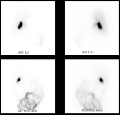
Description: Scan shows well-outlined liver with good perfusion of the tracer throughout the liver parenchyma. The gallbladder is visualized at the 10th minute of the study with the visualization of the intestine thereafter. Progressive accumulation of the tracer is noted in the gallbladder until the 60th minute of the study. Post-fatty meal images show partial contraction of the gallbladder with ejection fraction of 20% (normal > 30%). Significant amount of tracer is noted in the gut at the end of the study suggestive of no obstruction in the biliary tract.
Case Discussion
Laboratory investigations showed normal liver function tests (LFTs) and inflammatory markers.
No gallstones, features of acute cholecystitis or biliary obstruction were seen on the ultrasound and MRCP. Cholescintigraphy showed partially contracting gallbladder in response to the fatty meal and was suggestive of gallbladder dyskinesia with no obstruction in the biliary tract.
The patient was reassured and managed conservatively.


 Unable to process the form. Check for errors and try again.