
Presentation
Incidental finding on CT. Originally the study was targeted to vascular disease (endovascular aortic aneurysm repair infection).
Patient Data
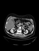

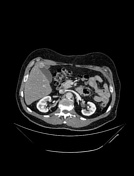

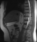
2 cm well-defined rounded image located in the fundus portion of the gallbladder, in the venous phase there is a partial enhancement at the periphery.
Partial pancreatectomy.
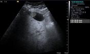
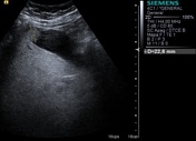
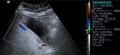

Abdominal US reveals a 2 cm mass located in the fundus, homogeneous, well-demarcated, non-mobile and avascular, suggestive of a polyp.

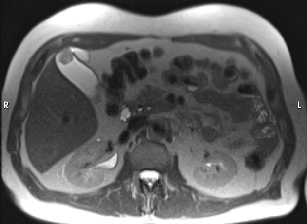
Axial and sagittal projection in T2 reveals a well-marginated lesion inside the gallbladder, of villus morphology, approximately 18 x 17 mm in size.
No evidence of wall thickening or stones in the gallbladder.
Partial pancreatectomy changes.
No other findings.
Case Discussion
Cholecystectomy was performed. The histopathology study revealed an adenomyomatosis with low-grade dysplasia without evidence of malignancy.


 Unable to process the form. Check for errors and try again.