
Presentation
Brown stained vomitus, palpable small bowel loops. Bowel obstruction?
Patient Data
Age: 85 years
Gender: Female
From the case:
Gallstone ileus
Download
Info
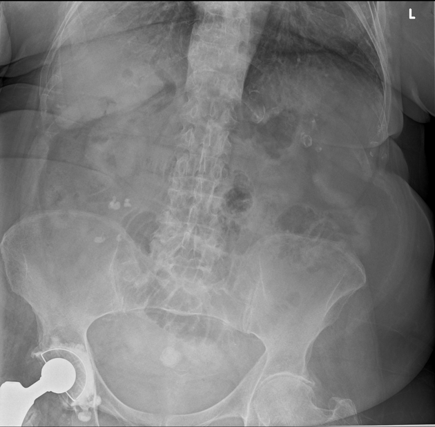
Prominent small bowel loops centrally in the pelvis.
Air in the CBD.
Calcified density in the midline of the pelvis.
From the case:
Gallstone ileus

Download
Info
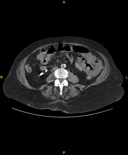
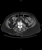
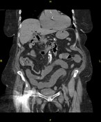
Pneumobilia in the left hepatic lobe. Cholecystoduodenal fistula.
1.5 cm calculus in the terminal ileum approximately 5-10 cm from the ileocaecal valve. The small bowel loops proximal to this are distended with fluid.
Kidneys, adrenals, spleen and pancreas appear normal.
Case Discussion
The plain radiograph demonstrates the Rigler triad of:
- pneumobilia
- small bowel obstruction
- ectopic calcified gallstone (in the small bowel)
CT confirmed the plain radiograph suspicion of gallstone ileus.
The plain radiograph is a classic fellowship viva case.


 Unable to process the form. Check for errors and try again.