
Presentation
Right suprarenal mass discovered on sonography.
Patient Data


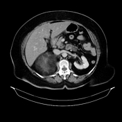
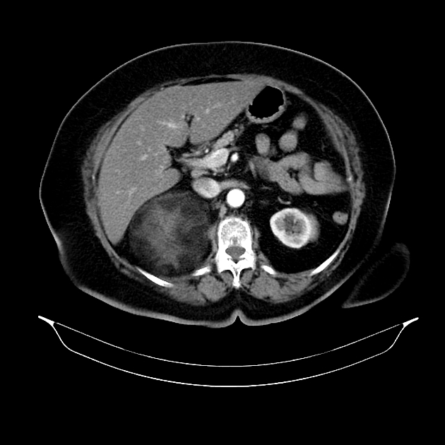
A large mass in the retroperitonium on the superior and lateral aspects of right kidney. The kidney was displaced inferiorly. Superiorly, the mass abuts the posterior aspect of the liver and narrows the inferior vena cava.
The tumor was well-circumscribed and there is no invasion of surrounding structures. The mass showed predominantly fatty regions interspersed with areas of higher attenuation inside. Density on CT varied between –60 Hounsfield units (HU) peripherally to –40 HU in the center. The mass is measuring 12.1 cm in its maximal diameter.
Two right hepatic lobe peripheral sub-capsular hepatic focal lesions are seen showing irregular enhancement on portal venous phase, and more homogeneous enhancement on the delayed phase, in keeping with hemangiomata.
Case Discussion
- location and attenuation of the mass on CT are compatible wtih right adrenal myelolipoma.
- adrenal myelolipomas are rare benign tumors of the adrenal gland with varying proportions of hematopoietic and adipose tissue.These lesions rarely measure more than five cm in diameter, although giant tumors have been occasionally reported.
- giant adrenal myelolipomas may be asymptomatic, or because of their large size may lead to dragging abdominal pain, an abdominal mass, compression of neighboring organs and even acute intratumoral or retroperitoneal hemorrhage
- the main differential diagnosis will be liposarcoma based on the large size of the tumor and presence of adipose components.
- giant adrenal myelolipomas are usually treated by simple adrenalectomy and completely curable. Hence an awareness of this rare adrenal entity and its correct diagnosis is important.


 Unable to process the form. Check for errors and try again.