
Presentation
Known giant cell arteritis with painful left temple. On prednisolone. Now developing right leg claudication.
Patient Data
Right lower limb arteries


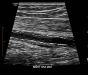
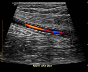

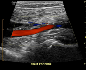
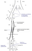
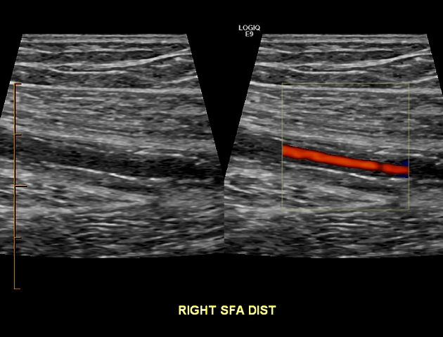
Findings similar to that seen in the superficial temporal artery with circumferential hypoechoic wall thickening (halo) and increased flow velocities in the distal superficial femoral artery. Proximal and distal arteries are essentially normal.
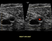
Lt superficial temporal artery

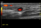
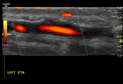
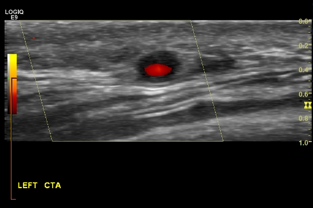
Typical pattern of circumferential hypoechoic wall thickening involving the painful left superficial temporal artery.
Case Discussion
Although giant cell arteritis (GCA) typically involves the extra-cranial arteries especially the superficial temporal, it can involve intracranial arteries especially the retinal as well as larger arteries including the aorta and its branches. The typical ultrasonic appearance is a cuff of hypoechoic tissue causing smooth luminal narrowing and increased flow velocities on duplex imaging.


 Unable to process the form. Check for errors and try again.