
Presentation
Complains of left wrist pain post trauma.
Patient Data
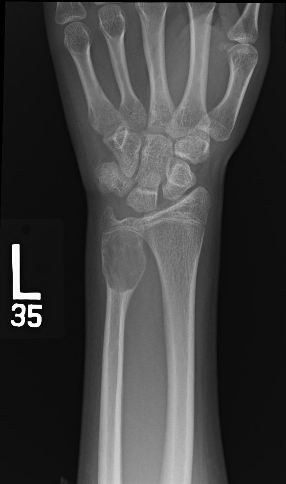
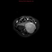
An expansile lucent bone lesion with narrow zone of transition arising from the metaphysis of the distal ulna bone.
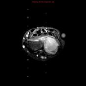

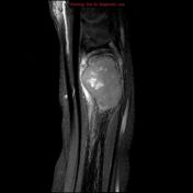
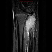

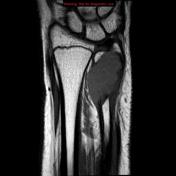
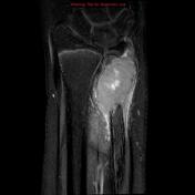
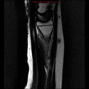
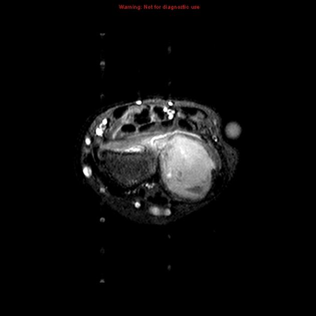
And expansile bone lesion arising from the distal ulna metaphysis, which demonstrates low signal in T1 and heterogeneous signal in T2. Foci of low signal in T2 representative of hemosiderin or fibrosis. Post gadolinium acquisition demonstrates enhancement.
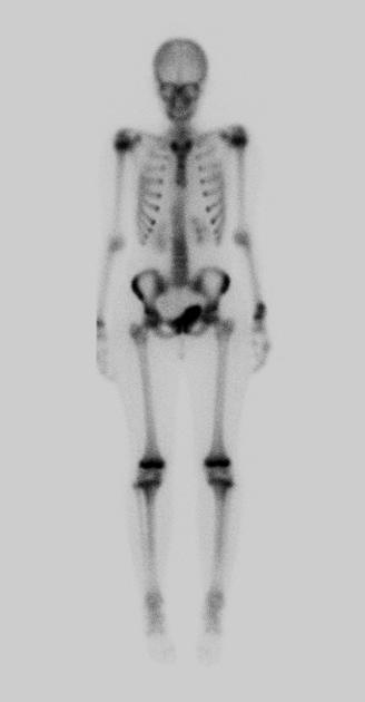
Increase uptake seen at the left wrist corresponds to the lesion seen in the distal ulna bone. Delayed acquisition of the left arm further demonstrates a central photopenic region (doughnut sign).
Case Discussion
Pathologically proven giant cell tumor. What is peculiar about this case is that the growth plates are still open!


 Unable to process the form. Check for errors and try again.