
Presentation
Incidental finding of a liver lesion during a routine abdominal ultrasound examination.
Patient Data
Age: 30 years
Gender: Female
From the case:
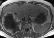
Giant liver hemangioma

Download
Info

Large T2 fat sat hyperintense lesion with smooth and sharp borders in the right hepatic lobe. After contrast administration, the lesion shows centripetal nodular rim enhancement.
Case Discussion
Typical MRI findings in a giant liver hemangioma are (among others) marked hyperintensity on long TE images and nodular rim enhancement after contrast administration.


 Unable to process the form. Check for errors and try again.