
Presentation
Chest pain.
Patient Data
Age: 85 years
Gender: Female
From the case:
Giant pericardial cyst


Download
Info

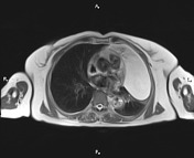
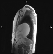
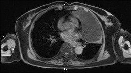
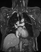
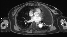
A 130×70×110 mm thin-walled non-enhancing cystic lesion is present at the anteroinferior aspect of the left hemithorax in close contact with the heart, causing mild adjacent parenchymal collapse. Trace pleural fluid is present bilaterally.
Case Discussion
Features on MRI are most consistent with a giant pericardial cyst, which is an uncommon benign congenital anomaly of the anterior and middle mediastinum. They are most commonly found on the right side, in particular the right anterior cardiophrenic angle, but can be found almost anywhere adjacent to the heart.


 Unable to process the form. Check for errors and try again.