
Presentation
Bilateral high myopia, esodeviation of the eyes and a history of left eye high intraocular pressure.
Patient Data
Age: 40 years
Gender: Female
From the case:
Glaucoma and chorioretinal coloboma

Download
Info

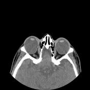
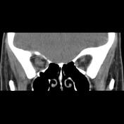
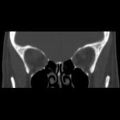
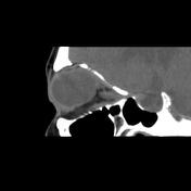
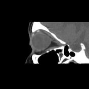
Both eyes are egg-shaped with a large AP length up to 35 mm and a thin posterior uveo-scleral layer. Esodeviation of the eyes more prominent on right side. Small caliber left optic nerve and implanted aqueous tube shunt in superotemporal aspect of the left orbital cavity are seen.
Case Discussion
The case illustrates the non-contrast MDCT features of the chorioretinal coloboma and implanted aqueous tube shunt for controlling glaucoma 1-3.


 Unable to process the form. Check for errors and try again.