
Presentation
Seizures.
Patient Data
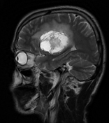


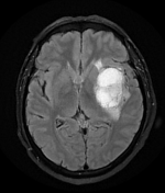
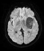
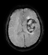
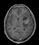
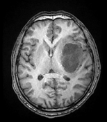
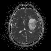
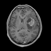
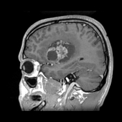
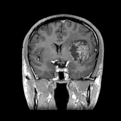
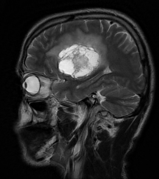
Left deep insular fairly defined complex space-occupying lesion. It measures about 4.0 x 5.6 x 4.8 cm along its max TS, AP and CC dimensions. It elicits heterogeneous signal with cystic and solid components. It shows areas of blooming on SWI suggestive of hemorrhage/calcifications. No diffusion restriction. It shows heterogenous enhancement. It's surrounded by vasogenic edema and exerts positive mass effect on the left lateral ventricle.
Differential possibilities include:
metastatic lesion
grade 4 diffuse astrocytoma/glioblastoma
parenchymal ependymoma
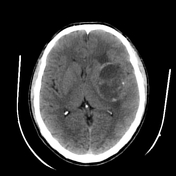
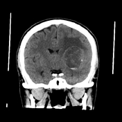
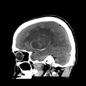
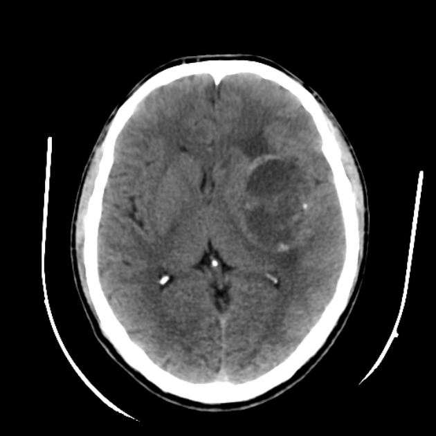
The lesion appears complex on non contrast CT scan with hyperdense hemorrhagic areas and small calcific foci.
The patient went on to have a biopsy.
Histology
Sections revealed brain tissue infiltrated by malignant astrocytic tumor compromised of malignant astrocytes with pleomorphic nuclei, increased nuclear-cytoplasmic ratio, prominent nucleoli and frequent mitosis. Vascular proliferation was detected in the form of small blood vessel proliferation and endothelial proliferation with occasional glomeruloid morphology. Focal necrosis was observed.
Final diagnosis
WHO CNS grade 4 astrocytic tumor.
Immunostain:
P53: negative staining
IDH1;Negative staining
GFAP:Diffuse positive staining
K167:Proliferation index 8%
Case Discussion
This is a highly aggressive complex cystic solid lesion. Differential possibilities according to the relatively young age of the patient included:
grade 4 diffuse astrocytic tumor, either an astrocytoma or glioblastoma
parenchymal extraventricular ependymoma
pilocytic astrocytoma with piloid features in NF patients
metastatic lesion
The immunostain was negative for IDH 1 mutation, however, for the diagnosis of IDH wild type in this young age (<55 years) the patient must have sequencing which is unfortunately not made for this patient. This makes the diagnosis (Glioblastoma NOS).


 Unable to process the form. Check for errors and try again.