
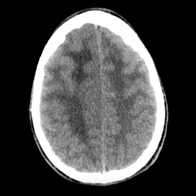
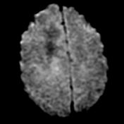
A single image from a non-contrast CT demonstrating widespread abnormality involving both hemispheres. There is white matter hypodensity, however, the cortex also appears thickened.

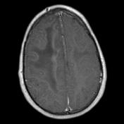
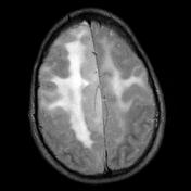
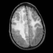
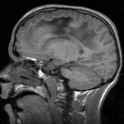
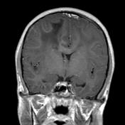

MRI confirms bilateral abnormalities with high T2 signal within the white matter and extensive thickening of the cortex most easily seen along the medial surface of the right frontal lobe. Changes involve at least three lobes and are consistent with gliomatosis cerebri.
Case Discussion
Diagnosis
Right frontal cortex and white matter: Diffusely infiltrating astrocytoma with a pattern consistent with gliomatosis cerebri (WHO Grade III).
Histology
The sections show a moderately cellular astrocytoma. The astrocytes are unevenly distributed and they show enlarged hyperchromatic nuclei with mild pleomorphism. The tumor cells extend into the cerebral cortex with perineuronal satellitosis. One mitosis is seen. There is no evidence of endothelial cell hyperplasia or necrosis. The neuropil is focally edematous and vacuolated. Many cells are p53 positive. The Ki-67 index is about 1%.
The features are those of diffusely infiltrating astrocytoma with a pattern consistent with gliomatosis cerebri. Most of these lesions are regarded as grade III.
Importantly, whereas gliomatosis was previously considered a distinct entity, since the 2016 update to the WHO classification of CNS tumors it is now merely thought of as a growth pattern.


 Unable to process the form. Check for errors and try again.