
Presentation
Headache, gait ataxia, and vomiting for more than 3 months.
Patient Data


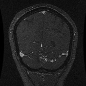
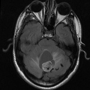
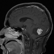
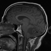
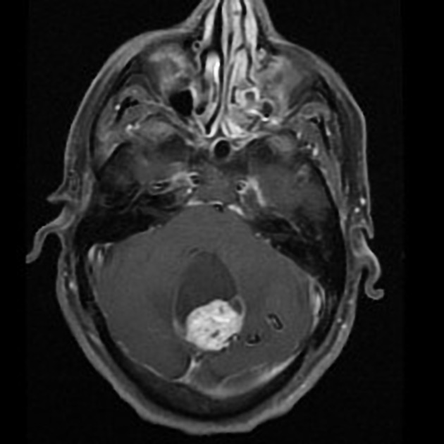
A vividly enhancing mass centered on the cerebellar vermis has an anterior cystic component. No convincing enhancement of the cyst wall. Some vasogenic edema in the cerebellar hemispheres. Prominent serpiginous flow voids within and adjacent to the nodule are seen confirmed to be abnormal draining veins to the left transverse sinus on post-contrast MRV.
The fourth ventricle is distorted by the cysts without, however, significant supratentorial hydrocephalus.
The features are highly consistent with hemangioblastoma.

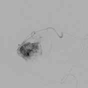
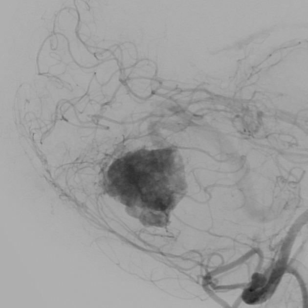
Angiography study at the time of preoperative embolization shows muliple enlarged feeding arteries primarily the superior cerebellar artery but also inferiorly from branches of the PICA and/or meningeal arteries. There appears to be an aneurysm at the origin of this enlarged vessel.
Dense tumor blush with early venous drainage superolaterally to the transverse sinus.
Case Discussion
The lesion was resected and was confirmed to be a hemangioblastoma.


 Unable to process the form. Check for errors and try again.