
Presentation
Patient presenting with altered consciousness and headache.
Patient Data


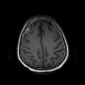
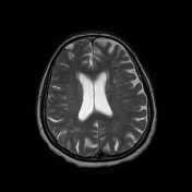
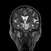
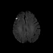
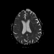
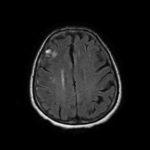
There are multiple cortical based T1w, T2w and FLAIR hyperintense lesions noted in the right frontal region, right parietal region and left side of the cerebellum.
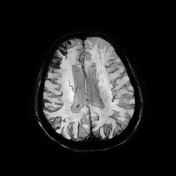
Lesions show a T2w hypointense rim. There is blooming in SWI sequences.
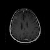
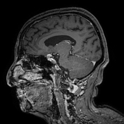
Diffusion restriction is noted and there is post contrast enhancement.
Prominent lesion seen in the R/frontal region measures 1.8 x 1.5 cm.
Some of the lesions abut the dura.
Sulci and vetnricular system are prominent suggesting cerebral atrophy.
Grey and white matter demarcation is preserved.
Basal ganglia appear normal.
Case Discussion
Appearances are in keeping with multiple hemorrhagic brain metastasis involving supratentorial and infratentorial compartments.
This patient was previously diagnosed with breast carcinoma and underwent mastectomy a few years ago. Hemorrhagic brain metastasis are compatible with the known breast cancer.
Other causes of hemorrhagic brain metastasis can be known by the mnemonic MRCT BB:
- melanoma.
- renal cell carcinoma
- choriocarcinoma
- thyroid cancer
- breast cancer
- bronchus/lung cancer
Patient was referred for oncological management.


 Unable to process the form. Check for errors and try again.