
Presentation
No history available
Patient Data
Age: Adult
Gender: Female

Download
Info
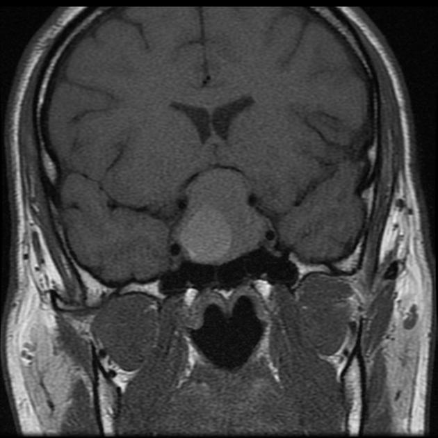
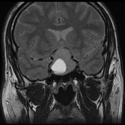
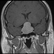
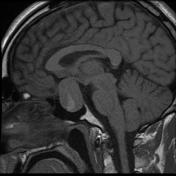
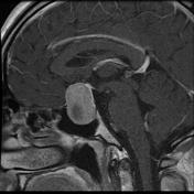
MRI of the pituitary demonstrates a large mass expanding the sella and extending into the suprasellar cistern compressing the optic chiasm. Within the right side of the mass a region of intrinsic high T1 and T2 signal is demonstrated (high T1 signal material is seen layering posteriorly) consistent with subacute blood products (extracellular methemoglobin).
Case Discussion
This case illustrates appearances consistent with hemorrhage into a pituitary macroadenoma. It would be interesting to know if the patient presented with sudden headache/visual loss to suggest classic pituitary apoplexy.


 Unable to process the form. Check for errors and try again.