
Presentation
History of seizure, acute RUQ pain
Patient Data


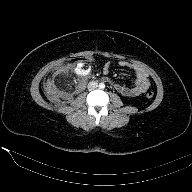
Multiple variable size fat density renal masses with enlarged vasculature are visible bilaterally in favor of bilateral renal angiomyolipomas. A hematoma is seen in the right perinephric space, deviating the right kidney anteromedially. The hematoma is due to hemorrhage of the largest angiomyolipoma of the right kidney. The hematoma extends to the combined retroperitoneal fascia, and mild to moderate free fluid with a fluid-fluid level is seen in the pelvis. Evidence of the previous cholecystectomy is seen.
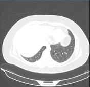
On lower thoracic CT images, several small pulmonary cysts are visible with LAM's possibility in the context of tuberous sclerosis.
Case Discussion
The patient likely is a case of tuberous sclerosis considering history of seizure and pulmonary cysts but the brain MRI was not available. She underwent trans-arterial embolization of the hemorrhagic mass.


 Unable to process the form. Check for errors and try again.