
Presentation
RUQ tenderness and fever.
Patient Data
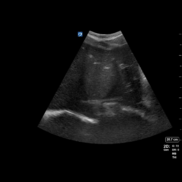
The liver is enlarged, measuring 173 mm. Lobular hypoechoic lesion within hepatic segment 6/7, measuring 94 x 80 x 71 mm with irregular internal septations. Vascularity cannot be accurately assessed with this ultrasound machine. No other hepatic lesions. The portal vein is 11 mm in caliber and has normal antegrade flow. The hepatic veins are patent.
The gallbladder is thin-walled (2 mm) and does not contain stones or biliary sludge. No intra- or extrahepatic biliary tree dilation. The common bile duct measures 4 mm in diameter.
The right and left kidneys are normal, and measure 141 mm and 149 mm respectively. No hydronephrosis.
The spleen measures on the upper limit of normal (132 mm).
No intra-abdominal free fluid. Pancreas and aorta are unremarkable.
IMPRESSION
Single complex lesion within hepatic segment 6/7, which is favored to represent an abscess in the given clinical context.
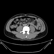

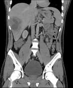
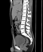
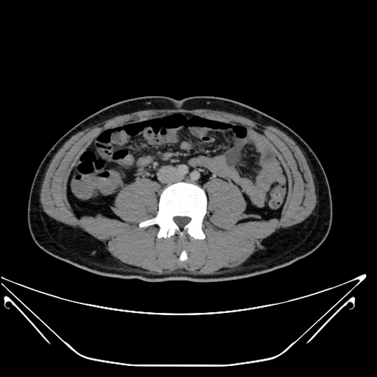
Lobular hypoechoic lesion within segment 6/7 of the liver, measuring approximately 95 x 61 x 84 mm (AP x TV x CC). Centrally the density measures 20 HU and contains multiple thick internal septations. Mild periportal edema present. The biliary tree is not dilated.
No intra-abdominal free fluid or gas. No peripherally enhancing collection identified elsewhere.
The spleen, pancreas, adrenal glands, and kidneys are normal. The small and large bowel are grossly normal with no focal bowel wall thickening.
No para-aortic, para-iliac, or inguinal lymphadenopathy. The abdominal aorta is not dilated. No filling defects within the portal or splenic veins.
The visible lung bases are clear. No concerning osseous lesion.
IMPRESSION
Right hepatic complex lesion is consistent with the previous ultrasound findings of a liver abscess. No vascular thrombosis or other complication evident.
Case Discussion
Pathology
Aspiration of liver abscess
MACROSCOPIC: Less than 1ml of thick, brown fluid received. 2 slides prepared.
MICROSCOPIC: The specimen contains leukocytes (predominantly neutrophils) and erythrocytes in the background of debris.
SUMMARY Aspiration of liver abscess: consistent with an abscess. No malignant cells found.
MCS
Gram Stain: Leukocytes 3+, Gram negative rods resembling Anaerobes 2+
Culture: Fusobacterium nucleatum 2+
Beta-lactamase Negative.
PARASITOLOGY
No ova, cysts or parasites seen.
The patient was treated with percutaneus drainage and recovered well.


 Unable to process the form. Check for errors and try again.