
Presentation
Right upper quadrant pain. Mildly elevated CRP and gamma GT.
Patient Data
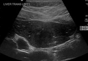
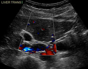

Multiple indistinct heterogenous liver lesions with minor internal vascularity. Images show a 2.6 cm lesion.
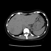


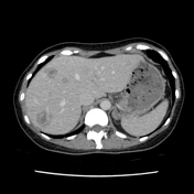
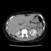

Multiple liver lesions demonstrating target appearance. Some are coalescent. Portal vein branches enter the periphery of some lesions (lollipop sign). Transient enhancement differences associated with some lesions.

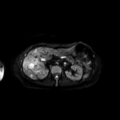
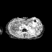
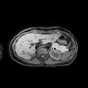
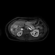
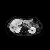

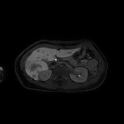
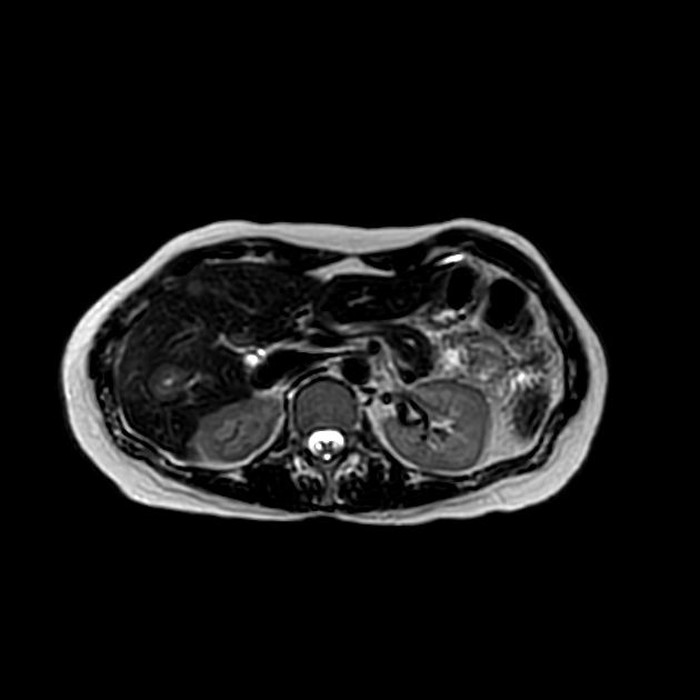
T2 DWI ADC T1 FS & Post Primovist
Twenty or more discrete and coalescent liver lesions demonstrating target pattern with rim enhancement on arterial phase imaging corresponding to peripheral diffusion restriction.
Lesion-associated transient enhancement differences.
Case Discussion
Histology from liver biopsy:
Sections show multiple cores of liver parenchyma in which the lobules and portal tracts are disrupted by numerous atypical epithelioid cells with fibrosis and scattered lymphocytes and neutrophils. Although the normal lobular structures can be highlighted by reticular fibers, neoplastic cells are intermingling with non-neoplastic hepatocytes, which are present as either single cells or two cells per plate. The neoplastic cells appear to be present within the vascular channels and some of them contain intracytoplasmic lumen in which red blood cells can be seen occasionally. Immunohistochemical studies were performed. These neoplastic cells are positive for ERG, CD31, and CD34, but negative for Pancytokeratin, CD68, Sox10, Hep Par1, Pax5, CD3, and EBER (ish). CD68 highlights Kupffer cell siderosis.
The overall features are consistent with epithelioid hemangioendothelioma.


 Unable to process the form. Check for errors and try again.