
Presentation
Abdominal pain and non-bilious vomiting for one day. Palpable right subcostal mass. History of omphalocele repair in the neonatal period.
Patient Data


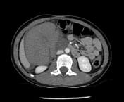
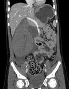
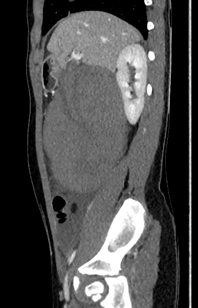
Anomalous position of the left hepatic lobe (LHL), below the right hepatic lobe (RHL). The LHL shows a ‘teardrop’ morphology with the vascular pedicle directed superiorly. Normal position, attenuation, and contrast enhancement of the RHL. Between the RHL and the stomach, a small segment of the liver is detected, very atrophic (lamellar morphology), with preserved attenuation and contrast enhancement.
Signs of ischemia of the LHL (absence of parenchymal contrast uptake). Vascular structures, such as the left portal vein, are non-opacificated. The left hepatic artery is filiform, almost absent; and the left hepatic vein sharpens towards the ischemic lobe until it is no longer visible. It causes compression of the pylorus and duodenum, leading to dilation of the gastric chamber. Fat stranding around the lobe and free fluid are also seen.
Abnormally positioned gallbladder (medially to the LHL), with thickened edematous wall.
Findings compatible with torsion of the LHL.
Urgent exploratory laparotomy confirmed the radiological findings: an abnormal almost separate hepatic lobe was torsed around the vascular pedicle and was necrotic. After detorsion, the parenchyma remained non-viable and was removed.
The complex anomalous liver anatomy leads to uncertainty regarding the segmental origin of the torsed lobe, probably a hypertrophic segment 4. Segments 2 and 3 probably account for the atrophic lamellar liver segment between the stomach and LHL. Other possibilities are atrophic LHL or an accessory lobe.
Case Discussion
Anatomically abnormal livers are often asymptomatic and identified incidentally in the context of imaging studies or during surgery performed for other reasons. Torsion of a liver lobe or segment(s) is rare and has been described with accessory lobes, ectopic liver tissue, or post-surgical livers. Of the few cases documented in the literature, a small number had previously undergone omphalocele repair. This could be due to abnormal liver fixation, either congenital or post-surgical, with increased mobility predisposing to torsion.
Although late abdominal complications such as intestinal volvulus or bowel obstruction secondary to adhesions or bands are more frequent in children with omphalocele, a liver lobe torsion is a possible association and should be considered.
Case co-authors:
Tello Arnas, L. Radiology Department. Hospital Universitario La Paz, Madrid.
Sánchez Galán, A. Pediatric Surgery Department. Hospital Universitario La Paz, Madrid.
Jimenez Rodriguez, M. Pediatric Emergengy Department. Hospital Universitario La Paz, Madrid


 Unable to process the form. Check for errors and try again.