

Presentation
Left wrist injury - one month ago. Pain in the hypothenar region.
Patient Data
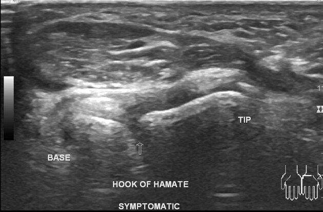
The symptomatic side shows a cortical breach involving the hook of the hamate. The Ulnar artery passing over the tip of the hook of the hamate is patent. A common variant accessory abductor digiti minimi muscle is present without ulnar nerve compression. The rest of the wrist examination was normal. The asymptomatic side hook of the hamate shows continuity.
Case Discussion
The patient fell on the ground while playing cricket. Initial frontal and lateral wrist radiographs were negative for a fracture (not uploaded, no copyright). He presented after one month for an ultrasound with a complaint of hypothenar region pain on local compression. The ultrasound shows a hook of hamate fracture.
The clinical examination of the hand revealed no skin changes/ local swelling in the hypothenar region. Finger movements and power were normal. No tingling along the ulnar nerve supply. No persistent pain which occurs when there is arterial thrombosis. The hook of the hamate fracture was suspected before holding the probe. During a normal wrist scan, only the tip of the hook of the hamate is seen. To see the entire length of the hook of the hamate, one has to slide the probe to the ulnar side in the short axis during volar scanning.


 Unable to process the form. Check for errors and try again.