
Ultrasound is a useful imaging modality for evaluation of the wrist, allowing high-resolution imaging of anatomy while simultaneously allowing dynamic evaluation of the joints, tendons, and ligaments.
Approach
There are multiple possible approaches to imaging the wrist with ultrasound. The exam is easily tailored to a specific painful area or set of differential diagnoses. A typical protocol is as follows 1:
Dorsal wrist
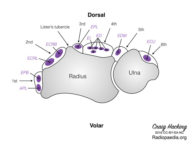
The hand is pronated. A transverse sweep across the dorsal wrist is sometimes helpful, identifying the extensor compartments 1-6. Lister's tubercle serves as a good landmark between compartments 2 and 3.
-
the hand is then put on its side, with the hypothenar eminence down. The first extensor compartment is then examined in short axis (abductor pollicis longus (APL) and extensor pollicis brevis (EPB)).
A septum may be present that splits the compartment.
-
while in this position evaluate radial artery and nerve
the artery is deep to the radial styloid
the nerve begins superficially to the compartment, and then proceed palmar to it
the hand is then pronated again. The second extensor compartment is then examined in short axis (extensor carpi radialis brevis and extensor carpi radialis longus (ECRB and ECRL).
the third extensor compartment is then examined in short axis (extensor pollicis longus tendon (EPL)). Particular attention should be paid to the area where the EPL crosses the ECRB and ECRL tendons.
the fourth and fifth extensor compartment is then examined in short axis (extensor digitorum communis, extensor indicis proprius, and extensor digiti minimi).
while over the 3rd-5th compartments, the scapholunate ligament can be assessed in short axis. Ulnar deviation of the wrist will stress the ligament and assess for patency.
the sixth extensor compartment is then examined in short axis and in long axis (extensor carpi ulnaris (ECU)).
in this position, the triangular fibrocartilage complex (TFCC) between the ulnar styloid and radius can be examined.
bringing the probe proximally, the distal radioulnar joint and midcarpal joints can be evaluated, in short and long axis, respectively.
Ventral wrist
The hand is supinated for examination.
evaluate the proximal carpal tunnel and distal carpal tunnel in short axis, with particular attention to the shape and echogenicity of the median nerve. The two bony structures outlining the sides of the carpal tunnel are the trapezium tubercle (radial) and hook of the hamate (ulnar). The median nerve can be examined in long axis as well.
the Guyon's canal and ulnar nerve can be assessed in short axis. The ulnar nerve is located more medially than the ulnar artery.
Pathology
A number of wrist abnormalities can be identified on ultrasound, including:
wrist effusion and/or synovial thickening (inflammatory/traumatic/septic)
erosions from inflammatory arthropathy
tendinosis, partial thickness tendon tear, full-thickness tendon tear
avulsion injuries
aneurysm/pseudoaneurysm
neuromas


 Unable to process the form. Check for errors and try again.