Presentation
Severe abdominal pain after percutaneous nephrolithotripsy (PCNL).
Patient Data
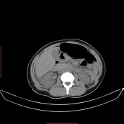

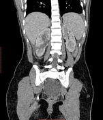

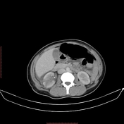

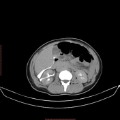

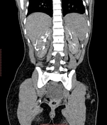

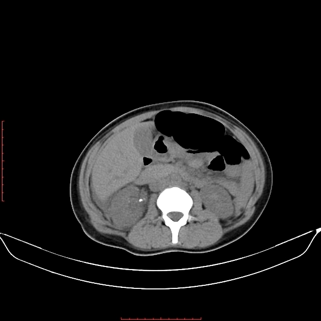
Lacerations of the inferior part of the right corticomedullary junction extending up to the collecting system associated with urinary extravasation in the hepatorenal pouch seen in the delayed images. Standing of the perinephric fat planes is seen. No evidence of active bleeding. A small muscle hematoma of the right posterior-lateral abdominal wall muscles and stranding of the subcutaneous fat is seen as well.
Retroaortic left renal vein (normal variant).
Few bilateral renal stones are seen dispersed within the different calyceal groups; the largest is seen in the right renal pelvis measuring about 0.8 cm in diameter with an average attenuation value of about 330 HU. No appreciable backpressure changes are seen.
Enlarged liver showing smooth outline and homogenous parenchymal texture with no focal lesion. No dilated intrahepatic biliary radicles.
Bulky spleen is noted. No focal lesions.
A small calcific focus is noted within the pancreatic head.
A moderate amount of free pelviabdominal collection.
Right adnexal cyst measuring about 5 cm in diameter, for US assessment.
IUCD is seen.
Case Discussion
CT is the best imaging modality for diagnosing the renal parenchymal injury. The delayed phase is a must to detect injury to the collecting system.
The American Association for the Surgery of Trauma (AAST) renal injury scale, most recently updated in 2018, is the most widely used grading system for renal trauma.
Severity is assessed according to the depth of renal parenchymal damage and involvement of the urinary collecting system and renal vessels into 5 grades.
Contrast-enhanced CT should be requested if the mechanism of injury or the clinical examination is suspicious for renal injury (e.g. RTA, lower rib fractures, flank ecchymosis, and any abdominal penetrating injury).


 Unable to process the form. Check for errors and try again.