
Presentation
Blurring of vision and reduced visual acuity for two weeks.
Patient Data


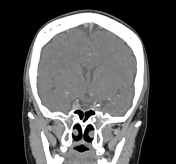
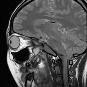
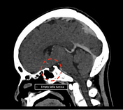
"Empty sella sign" where the normal pituitary gland height is lost within the sella turcica.
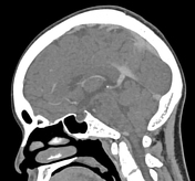
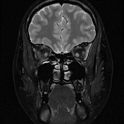
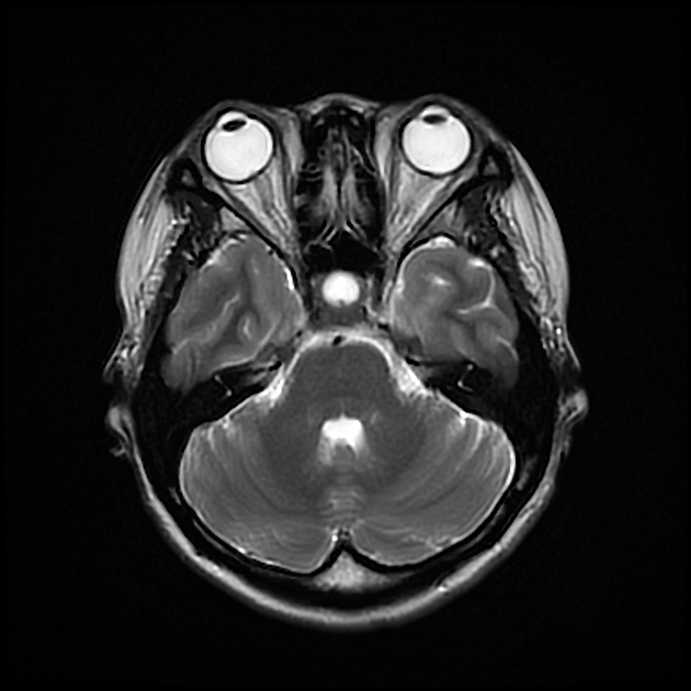
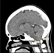
Acquired cerebellar tonsillar ectopia, where the bilateral cerebellar tonsils herniated below the McRae line by 5.5mm.
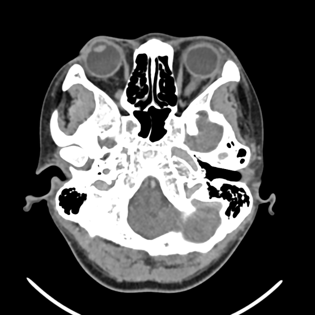
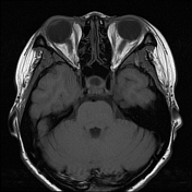
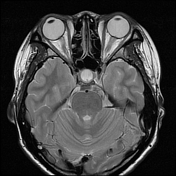
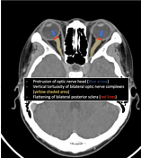
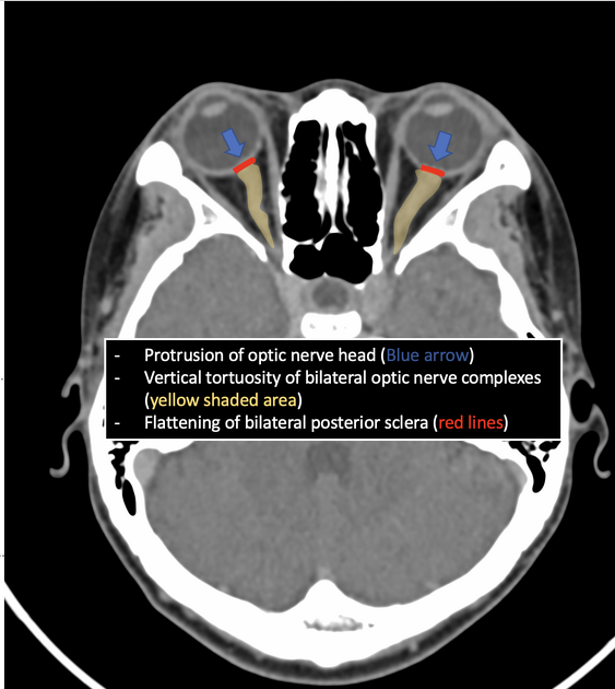
Flattening of posterior sclera bilaterally with mild protrusions of optic nerve heads. Mild tortuosity of the optic nerves.
CT venogram:
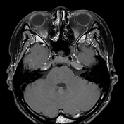
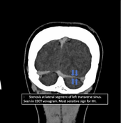
Cerebral venous sinuses and cavernous sinuses are well opacified. No abnormal filling defects to suggest thrombosis. Lateral segment of left transverse venous sinus appears to be stenotic comparing to the right side.

Similar findings as seen in previous CT scan :
Empty sella sign.
Flattening of posterior sclera at the optic discs
Protrusion of optic heads
Cerebellar tonsillar ectopia.
Annotated images showed the imaging features of idiopathic intracranial hypertension.
Case Discussion
Flattening of posterior sclera and protrusion of optic heads are CT imaging findings of papilledema. Constellation imaging findings of papilledema, empty sella turcica, transverse sinus stenosis and acquired tonsillar ectopia are suggestive of idiopathic intracranial hypertension.
Patient had lumbar puncture performed with abnormally high opening pressure (36mm Hg). Patient was treated with acetazolamide.
There are many other imaging features of idiopathic intracranial hypertension (such as enlarged Meckel caves, enlarged subarachnoid spaces around the optic nerves, enhancement of intraorbital optic nerves), where not all of them would be present at the same time.


 Unable to process the form. Check for errors and try again.