
Presentation
Initially shortness of breath. Then hemoptysis. Presentation during the height of COVID-19 pandemic.
Patient Data
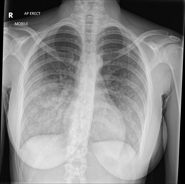
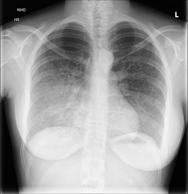
Diffuse symmetrical bilateral airspace opacification in both lungs in the mid and lower zones.
Heart size normal.
Normal mediastinal contours.
COVID-19 was considered a strong possibility.
PCR negative for COVID.


Bilateral breast prostheses.
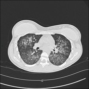
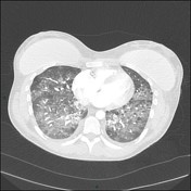
Widespread bilateral ground-glass change in both lungs with a lower lobe predominance.
No septal thickening. No pleural effusion. No mediastinal nodes.

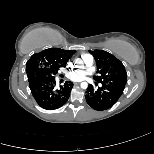
Bilateral breast prostheses.
No pulmonary embolus.
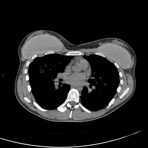
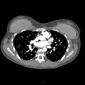
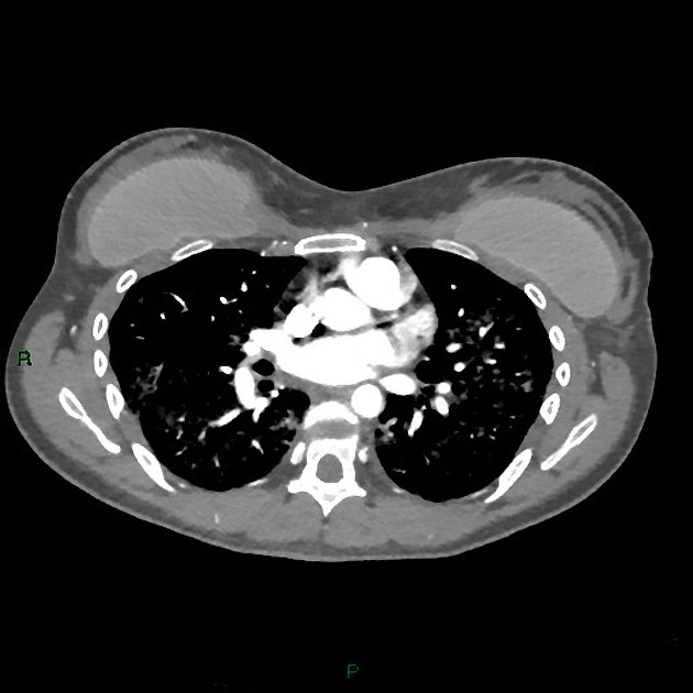
Diffuse, more confluent ground glass change/consolidation throughout both lungs, more pronounced in the lower lobes.
No mediastinal lymphadenopathy. No pleural effusions.
Bilateral airspace changes persist but with a right-sided predominance.
Slight regression in appearances overall.
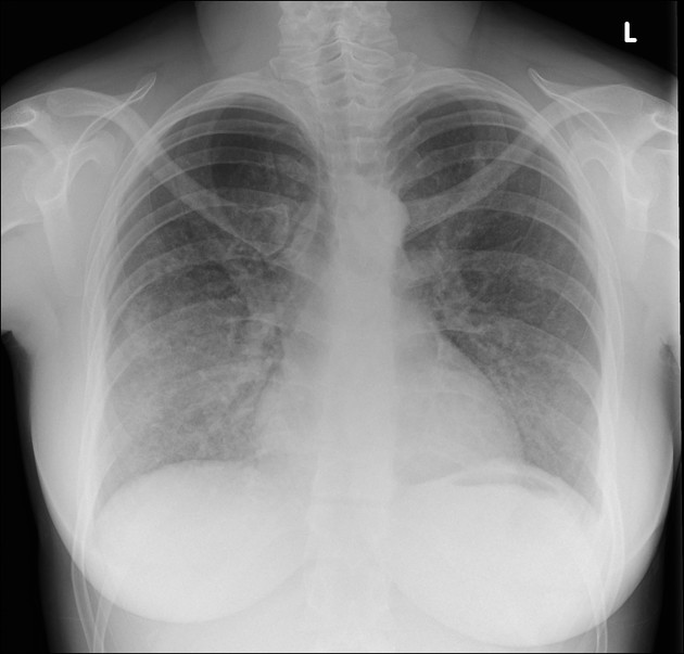
Bilateral airspace opacification, right more than left, similar to the prior film.
Heart size normal.
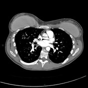
Bilateral breast prostheses.
No pulmonary embolus.
Diffuse bilateral ground-glass change throughout all lobes.
Case Discussion
Classical serial images of pulmonary hemorrhage.
Not unreasonably at the height of the COVID pandemic, the first chest radiograph was thought to relate to COVID not least as at that time the patient had no hemoptysis and was in the community.
The imaging has no specific features to identify a cause.
A transbronchial biopsy was undertaken and the results were in keeping with idiopathic pulmonary hemosiderosis.
This is a rare diagnosis and doesn't have any specific imaging appearances.


 Unable to process the form. Check for errors and try again.