
Presentation
Severe abdominal pain ? diverticulitis
Patient Data
Oral & IV C+ administered...


Oral & IV C+ administered as per a routine CT for diverticultis
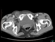
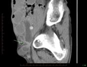
Gross dilatation of fluid-filled small bowel indicative of SBO down to the right groin where there is a hernial sac containing a loop of ileum. Note hernial sac is lateral to the pubic tubercle and compresses the femoral vein. Minimal ascetic free fluid. No portal or mesenteric venous gas. No area of bowel mucosal hypoenhancement seen.
Upper abdominal solid viscera are unremarkable.
Case Discussion
Femoral hernias can be difficult to differentiate from inguinal hernias on CT - key is the position of the incarcerated bowel relative to the pubic tubercle (PT) - indirect inguinal hernias start lateral and pass medial to the PT whereas femoral hernias have their neck medially but tend to pass lateral to the PT, extrinsically compressing the femoral vein so:
When interpreting a CT scan in a patient suspected of having a groin hernia, one may use the following algorithm: "When the hernia sac extends medial to the pubic tubercle, the diagnosis of inguinal hernia can be made with confidence. If the hernia sac is located lateral to the pubic tubercle, the presence of venous compression suggests the diagnosis of femoral hernia with a high probability"


 Unable to process the form. Check for errors and try again.