
Presentation
Severe epigastric pain of short time duration associated with nausea.
Patient Data
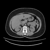

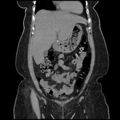
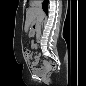
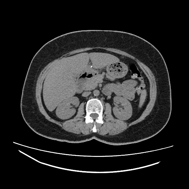
A hyperdense rod-like structure with smooth, well-defined edges is visualized piercing through the gastric antrum wall, extending from the lumen outward and abutting the left liver lobe consistent with gastrointestinal perforation.
However, there is no evidence of pneumoperitoneum or localized fluid collection.
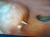
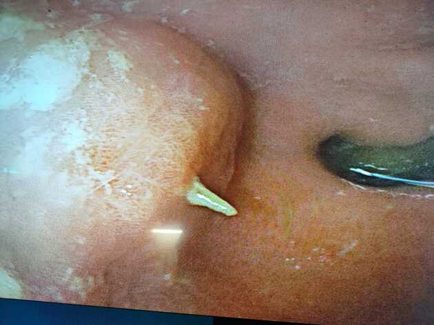
The upper endoscope shows a bone-like foreign body pierces the stomach wall.
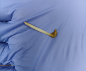
The ingested bone was removed by fibroendoscope.
Case Discussion
The initial clinical suspicion was acute cholecystitis, prompting an abdominal ultrasound, which returned unremarkable results.
After proper imaging, an urgent upper endoscopy was performed, and the foreign body was successfully removed. The object appeared to be a fishbone fragment, although the patient denied any recent history of eating fish or experiencing choking episodes.
Gastrointestinal perforation due to an ingested foreign body can lead to potential peritonitis and sepsis. Prompt diagnosis and treatment are crucial to manage this potentially life-threatening condition.


 Unable to process the form. Check for errors and try again.