
Presentation
Onset of pain in the throat while eating.
Patient Data

There is extensive soft tissue gas with the prevertebral space with associated swelling. A radiopaque foreign body can be seen inferior to the hyoid bone at the level of the C3 vertebral body.
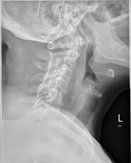
Arrow indicates location of the chicken bone.
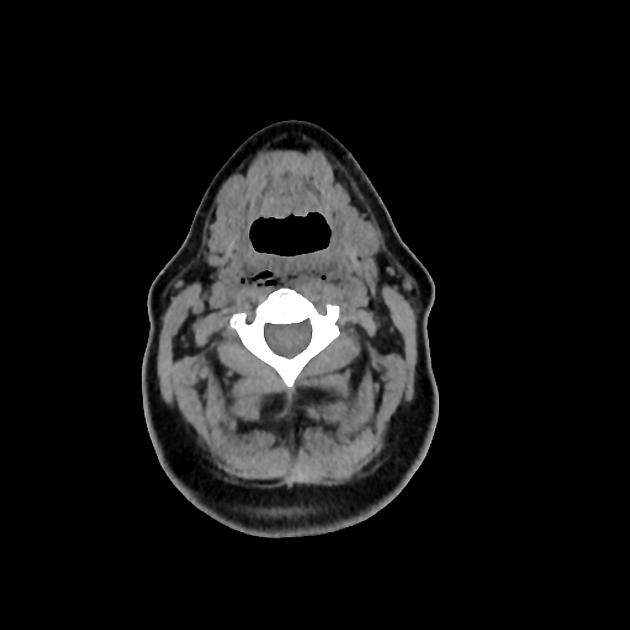
A foreign body can be seen piercing the prevertebral tissue, the tip of which lies in the posterior pharyngeal wall. The perforation has resulted in gas within the retropharyngeal and peripharyngeal spaces that extends to the base of the neck. Edema can also be seen involving the oropharynx.
Case Discussion
It is unusual to see imaging of a chicken bone, with fish bones being more common 1.
The bone passed prior to an attempt at removal and the patient was able to be discharged after a few days observation.


 Unable to process the form. Check for errors and try again.