
Presentation
Rule out pneumothorax
Patient Data
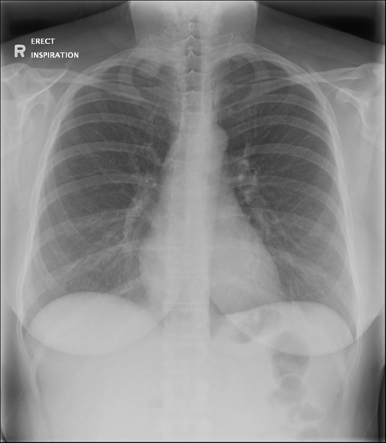
The radiograph was initially requested to rule out pneumothorax. There is no pneumothorax or other acute intrathoracic findings.
A rounded nodular opacity was incidentally found at the right thoracic apex, for which further assessment by CT was recommended.


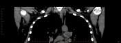
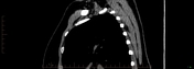
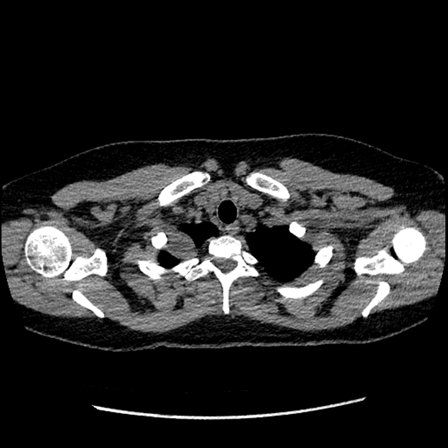
On CT, the well-defined right apical lesion is extrapleural, abutting the pleura and first rib, and seems to be associated with the T1 nerve root. Differential considerations include a peripheral nerve sheath tumor such as schwannoma versus a pleural based lesion such as solitary tumor of the pleura.
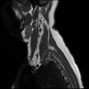
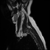
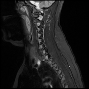
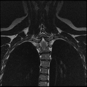
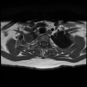
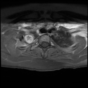
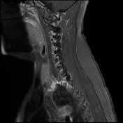

The right apical lesion can be seen arising eccentrically from the right first intercostal nerve on MRI. It is T1 iso- and heterogenously T2 hyperintense. There is avid post-contrast enhancement. The appearance is suggestive of a schwannoma of T1.
Case Discussion
Schwannomas are benign peripheral nerve sheath tumors. Our patient elected to have serial imaging follow-up for this mass, which remained unchanged in size and appearance on the two-year follow up scan.


 Unable to process the form. Check for errors and try again.