Presentation
Persistent locking of the left knee. History of trauma few years prior. ACL tear?
Patient Data
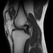

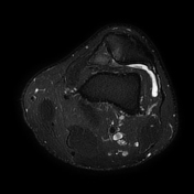

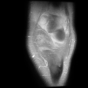

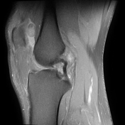

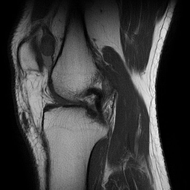
A 19 mm well-circumscribed mixed-signal loose body inside the medial aspect of the supra-patellar recess; bone erosion is visible in the adjacent patellar surface, but no evidence of invasion to the adjacent structures is seen; mild joint effusion. Two 6 and 5mm peri-cruciate ganglion cysts, posterior to the posterior cruciate ligament; the posterior cruciate ligament is intact. Low-grade sprain of the posterior aspect of the superficial medial collateral ligament fibers.
Low-grade old partial tear of the popliteus tendon, low-grade sprain of the arcuate popliteal ligament, and low-grade sprain of the femoral attachment of the fibular collateral ligament. Tri-compartmental degenerative changes; small ganglion cyst, associated with the medial head of the gastrocnemius muscle.
Case Discussion
The intra-articular loose bodies arise from a wide range of underlying causes. This case shows a relatively large intra-articular loose body in the supra-patellar recess, causing pressure erosion in the adjacent patellar surface. Regarding the size, morphological characteristics, and lack of evidence of any underlying fractures, osteochondral lesions, meniscal/ligamentous tears, and significant degenerative changes that could justify this loose body, a para-articular chondroma 1 would be the most probable diagnosis.


 Unable to process the form. Check for errors and try again.