
Presentation
Large head size and neurological impairment.
Patient Data
Age: 8 days
Gender: Male
Download
Info

CT without contrast demonstrates a very large hyperdense mass in the supratentorial compartment which has markedly heterogeneous density with components of fat, fluid and soft-tissue attenuation. Hyperdense material layers dependently in the occipital horns consistent with blood.
Marked supratentorial obstructive hydrocephalus with transependymal CSF permeation.


Download
Info
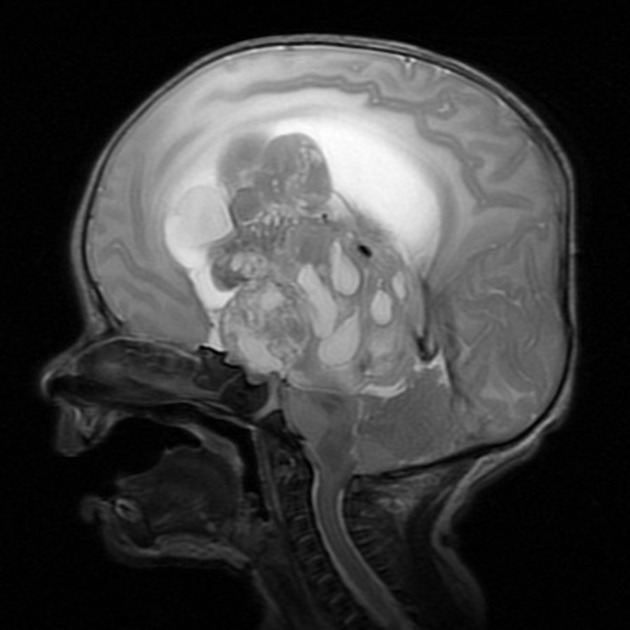
MRI confirms the findings, with a large heterogeneous mass with variable signal and blood layering both within cystic components of the mass as well as in the occipital horns.
Case Discussion
This child went on to have a craniotomy and debulking of the tumor, confirming the diagnosis of an immature intracranial teratoma.


 Unable to process the form. Check for errors and try again.