
Presentation
Left hemiparesis for the last 6 weeks.
Patient Data
Age: 16 years
Gender: Male
From the case:
Intracranial multifocal germinoma


Download
Info
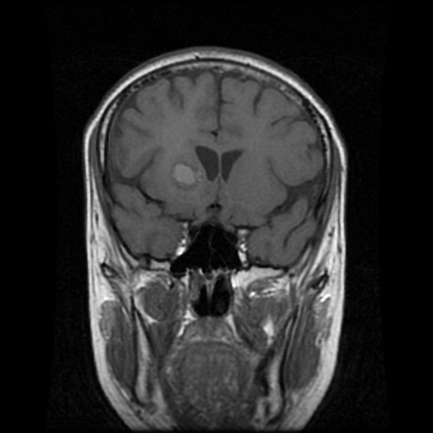
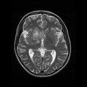
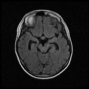
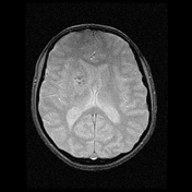
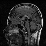
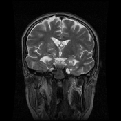
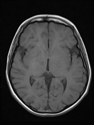
There are multiple abnormal space- occupying lesions located as below :
- ill-defined masses at right basal ganglia and right thalamus part of it showed suggestion of hemorrhagic signal changes or calcifications and containing cystic components with fluid-fluid level
- within 3rd ventricle measuring 1 X 1.2 X 1.5 cm resulting in mild hydrocephalus and associated with transependymal edema
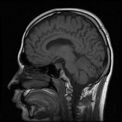
- sellar and suprasellar mass measuring about 1.1X1.8X2.5 cm
Note: artifacts are seen within CSF spaces at top of FLAIR sagittal images due to technical factors.
From the case:
Intracranial multifocal germinoma
Download
Info
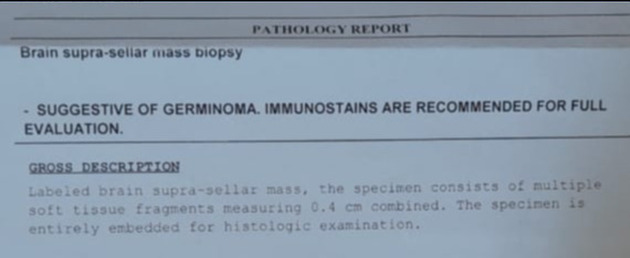
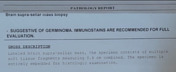
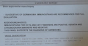
The patient underwent a supra-sellar mass biopsy and the final histopathological diagnosis is germinoma.
Immunostains for OCT4 and CD117 markers are positive. keratine and chromogranin markers are negative.
This panel supports the diagnosis of germinoma.
Case Discussion
Findings are consistent with multifocal germinoma located mainly at :
- right basal ganglia and right thalamus
- sellar and suprasellar cistern
- 3rd ventricle which is resulting in mild hydrocephalus


 Unable to process the form. Check for errors and try again.