
Presentation
Post fall.
Patient Data
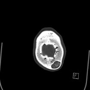

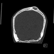
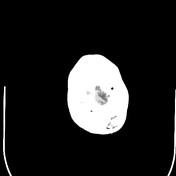
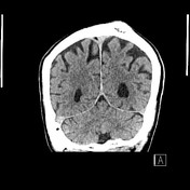

A slightly eccentric lesion measuring 2.7 x 2.0 x 1.3 cm is noted expanding the diploic space of the superior left parietal bone displaying a sclerotic margin and stippled internal matrix calcification. Bulging of the outer table without evidence of cortical breach. No associated soft tissue mass. The remainder of the calvarium is within normal limits.
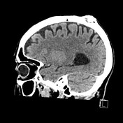
Mild parenchymal volume loss with ex vacuo dilation of the lateral and third ventricles. Minimal bilateral globus pallidus mineralization noted. The cerebral hemispheres, brainstem and posterior fossa are otherwise within normal limits without evidence of a focal intra-axial lesion. No acute intracranial haemorrage. No extra-axial fluid collections. Moderate bilateral cavernous carotid artery calcification.
Bilateral lens replacements. Minor bilateral maxillary sinus and ethmoid air cells mucosal thickening.
Case Discussion
A great example of non-destructive calvarial lesion most likely representing an intradiploic epidermoid cyst with sharply demarcated bony defects and zones of internal stippled calcifications.
Another differential diagnosis is skull vault hemangioma.


 Unable to process the form. Check for errors and try again.