
Presentation
Right arm painless swelling for many years. No pain. On examination, the swelling involves anterolateral side of the distal arm.
Patient Data
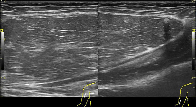
There is a well-defined, encapsulated, intramuscular lesion in the region of interest. It is in the mid-distal forearm on the anterior-lateral side and measures about 132 x 55 x 40 mm. The lesion is isoechoic to hyperechoic in comparison to the adjacent subcutaneous fat and hyperechoic to adjacent muscle. There are linear incomplete internal striations. Ther are few septa ( at least 2 septa, less than 2 mm thick ) in the lesion with a small (5 mm) calcification focus. At least two septa ( up to 2 mm thickness ) in the lesion. There is no non-fatty large solid area in the lesion. The lesion shows compressibility. A single small vessel traverses the lesion.
Case Discussion
An adult male presented with the right arm painless lesion which was present for many years. The ultrasound findings favor an intramuscular lipoma which was surgically excised.


 Unable to process the form. Check for errors and try again.