
Presentation
Knee pain.
Patient Data
Age: 55 years
Gender: Male
Download
Info
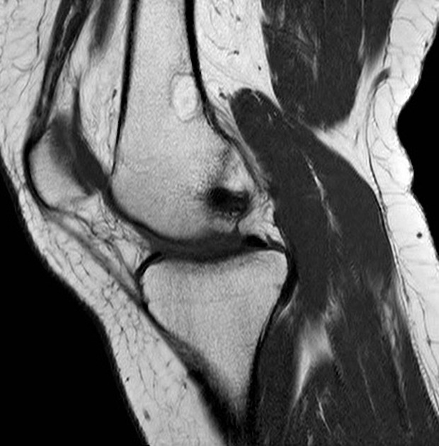
MRI shows a hyperintense T1 lesion located at the posterior distal diaphysis of the femur, compatible with intra-osseous lipoma. Not shown here, but the pattern should be: hyperintense T1 & hypointense T2 fat sat.


 Unable to process the form. Check for errors and try again.