
Presentation
Gradual onset right-sided proptosis and headache.
Patient Data


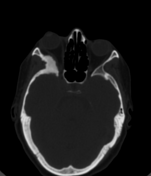
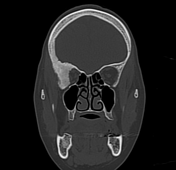
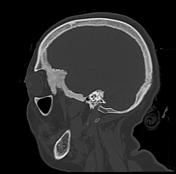
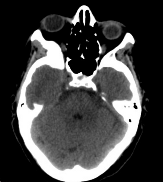
Focal bony expansion, remodeling, and hyperostosis with surface irregularity involving the greater wing of the right sphenoid bone, squamous part of the right temporal bone, right frontal bone, and superolateral orbital wall causing right exophthalmos, without obvious soft tissue component.
Case Discussion
Differential diagnosis includes fibrous dysplasia which shows expanded medullary bone with ground-glass attenuation; while intraosseous meningioma shows periosteal hyperostosis with surface irregularity.
This patient went on to surgery and histology confirmed the diagnosis of intraosseous meningioma.
PATHOLOGY REPORT:
GROSS:
Specimen fixed labeled with patient's name, consists of 2.0 gm, multiple brownish boney tissue pieces in aggregate measuring 3.0x1.5cm. Submitted all in one cassette.
MICROSCOPIC:
Sections reveal lobulated architecture, some meningothelial whorls composed of syncytial cells with indistinct membranes, eosinophilic cytoplasm, round uniform nuclei intranuclear pseudoinclusions are rare between trabecular bone.
The tumor cells are immunoreactive for EMA, Ki67 prolifertivity index is 1%.
DIAGNOSIS:
Right side of skull (sphenoid) bone: Intraosseous meningioma.


 Unable to process the form. Check for errors and try again.