
Presentation
Nasal obstruction and epistaxis. Previous paranasal sinus surgery a long time ago.
Patient Data


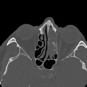
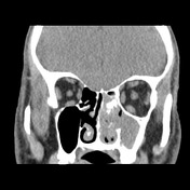
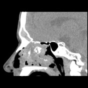
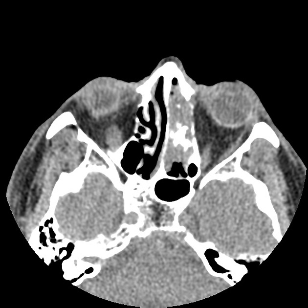
Soft tissue mass obliterating the left ethmoid cells, destroying the internal bony septa, and extending into the superior portions of the left nasal cavity and obliterating the left fronto ethmoidal recess.
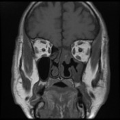
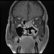
Prior left turbinectomy. Mucous thickening in the left maxillary sinus has patchy irregular enhancement and is also associated with signs of chronic hyperostosis and are most likely related with chronic sinus disease.

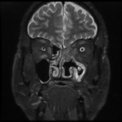
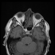

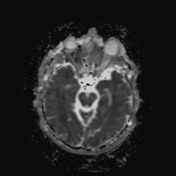
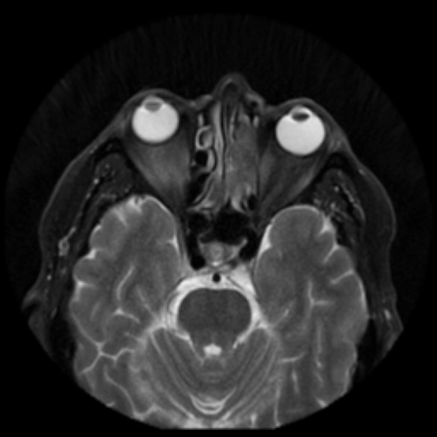
Soft tissue mass obliterating the left ethmoid cells, destroying the internal bony septa, and extending into the superior portions of the left nasal cavity and obliterating the left fronto ethmoidal recess. It shows to be isointense on T1 and slightly heterogeneously hyperintense on T2, with vivid contrast enhancement having a slightly "cerebriform" pattern. The left frontal sinus is fully filled by homogeneous fluid content, with only a thin smooth rim of mucosal enhancement. Similarly smooth but slightly thicker enhancing tissue abuts the inferior most aspect of the frontal sinus where the bony wall is attenuated. No evidence of invasion into the cranial vault.
MICROSCOPIC DESCRIPTION: 1. Sections show fragments of tissue with many bulbous nests of bland squamous epithelium within an edematous stroma. The surface of most fragments is bland squamous epithelium. There is a predominantly neutrophil infiltrate within the epithelium with some microabscesses. Keratinization is not prominent. There is some cytoplasmic clearing. Nuclear features are for the most part bland, with some areas showing reactive atypia. There is no evidence of malignancy.
DIAGNOSIS: Schiederian papilloma, inverted type.
Case Discussion
The main differentials for the lesions invading and destroying the ethmoidal sinuses on the left are sinonasal carcinoma and inverted papilloma.
The case was histologically proven to be an ethmoidal inverted papilloma, which shows to extend into the left frontoethmoidal recess, with fluid opacification of the left frontal sinus. No overt invasion into the frontal sinus, although its inferior wall is deficient. No evidence of intracranial invasion.


 Unable to process the form. Check for errors and try again.