
Presentation
Presenting with cough, investigated by a chest CT scan. Incidental single kidney found in the upper abdomen.
Patient Data
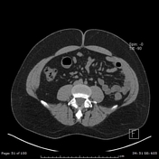

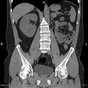


Empty left renal bed, consistent with left renal agenesis. The right kidney displays compensatory hypertrophy with a tiny cortical cyst arising from its lower pole. No right renal stones or hydronephrosis. The right ureter and urinary bladder are unremarkable.
Absent left SV (seminal vesicle). The left ductus deferens is absent compared to the normal R ductus. Normal right seminal vesicle.
Incidental small quantity of extraperitoneal fat in the left inguinal canal, (so-called ‘lipoma’).
Incidental ovoid hypoattenuating right adrenal lesion, likely representing a lipid-rich adenoma.


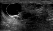
Left epididymal enlargement, demonstrating a speckled appearance secondary to tubular ectasia (tubular ectasia of the epididymis).
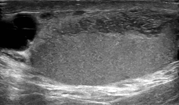
The mediastinum testis appears expanded, containing multiple enlarged anechoic tubular structures, consistent with tubular ectasia of the rete testis.
Irregular anechoic left epididymal cystic lesion, likely representing a small spermatocele.
This constellation of findings is consistent with obstruction of the ductus deferens.
Case Discussion
Agenesis of the seminal vesicles and ductus deferens is closely associated with urinary tract anomalies, usually renal agenesis, but other renal anomalies can also occur. Normal kidneys can be found in up to 9% of cases.
This is a case of unilateral seminal vesicle and ductus deferens agenesis, associated with agenesis of the ipsilateral kidney, with sonographic changes of left ductus deferens obstruction, manifested by tubular ectasia of the left epididymis and rete testis, associated with a small left spermatocele.


 Unable to process the form. Check for errors and try again.