
Presentation
Nine months old infant with clinically evident conjoined twins. Number (1) infant shows morphologically complete built and maturation. The second one is incomplete, with two legs, a rudimentary hand, and a penis with deformed scrotum [a total of 2 penises]. The twins are fused ventrally at the pelvic region of the complete one.
Patient Data

There’s duplication of both lower limb bones; namely: femoral, tibia, fibular, and foot bones.
- the laterally located lower limbs are of the complete infant; showing iliac, pubic and ischial ossification centers
- the abnormal lower limbs (of the incomplete twin) are seen medially placed, being flexed and directed upwards, showing bilateral ischium and pubic bones ossification centers, yet no iliac bones ossification center noted. Absent both femoral heads with acetabular dysplasia
A rudimentary single upper limb bones (radial ray, metacarpo-phalanges) are seen arising anteriorly at the pelvis with a segmented bizarre looking bony arch arising laterally from it.
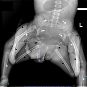
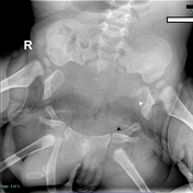
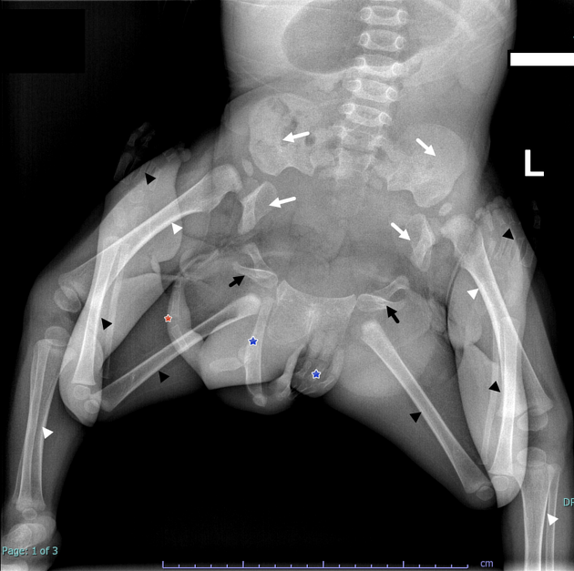
Fig 1. Pelvic x ray of the child, showing:
- white arrow heads: femur, tibia and fibula of the complete baby
- white arrows: ilium, pubis and ischium of the complete baby
- black arrow head: femur, tibia and fibula of the incomplete baby
- black arrows: pubis and ischium of the incomplete baby
- blue asterisk: single upper limb bones (radius, hand bones)
- red asterisk: abnormally segmented curved bone arising from the bones of upper limb
Fig 2: Spot on pelvis, showing: (white arrow head: pubis and ischium of the complete baby), (black arrow head: pubis of the incomplete one; being probably shared with the other one).

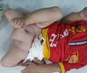
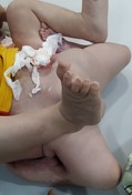
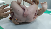

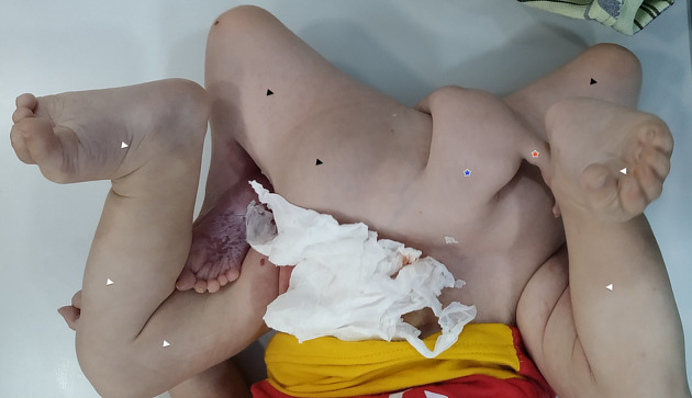
Photos of the baby:
- white arrow heads: lower limbs of the complete baby
- black arrow head: lower limbs of the incomplete baby
- blue asterisk: the abnormally shaped single arm
- red asterisk: the abnormally segmented curved structure arising from the arm
- black arrow head: lower limbs of the incomplete baby
- blue asterisk: the abnormally shaped single arm
- orange arrowhead: two penises of the complete and incomplete babies
Case Discussion
Conjoined twins are a type of mono-zygotic twin in which the embryonic division fails to occur during days 13 to 15 post-conception 1,2. They are an infrequent incident without an exact prevalence, although prevalence is reported to vary from 1 in 50000 to 1 in 100000, showing a higher incidence in females in Asia and Africa 3-5. The most widespread classification of conjoined twins is based on the place where they are united.
This 9-month-old patient was recently presented in July 2020. The clinical history as per parents was quite vague. They stated they’re seeking surgical operation for their baby. Only pelvis X ray was requested and performed. Unfortunately, antenatal care wasn’t available for the patient’s mother.
The conjoined twins are usually discovered during prenatal ultrasound 3. There’s no modality of choice, nevertheless a combination of ultrasound and MRI may be of a good benefit for prenatal diagnosis 6. Postnatal imaging aims mainly to delineate the complexity of fusion and its site and if there’s possibility of (shared) organs, as well as vasculature and innervation.
The prognosis and outcomes of the corrective surgeries depend on many factors, like degree of fusion, site of fusion, time of the operation postnatally and general condition of the neonate or child. Imaging plays a pivotal role in surgery planning, especially combination of various modalities and images reconstructions 6.
Take home message/teaching points
conjoined twins are a rare occasion
the ischiopagus type (ventral pelvic fusion) is very rare type with about 5% prevalence
it’s classified according to the most prominent site of interconnection
Final diagnosis
(conjoined twin) with ventral pelvic fusion (ischiopagus).
*Consented from parents for anonymized photograph and publish.
Case co-author:
Sherif Mohsen Shalaby
Mohamed A. Shanah
Ahmed Abuelnoor
Hala Sayed


 Unable to process the form. Check for errors and try again.