
Presentation
Left eye proptosis.
Patient Data
Age: 10 years
Gender: Female
From the case:
Juvenile ossifying fibroma


Download
Info
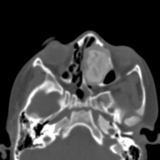
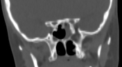
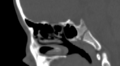
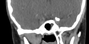
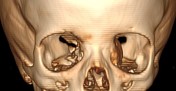
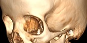
There is a well-defined lesion with bone density in the left ethmoidal and left frontal sinuses. Medially the lesion is not separable from the nasal septum. Laterally the lesion bulges into the left orbital cavity however no intraocular extension of the lesion is noted. Mild proptosis of the left eye is noted.
Case Discussion
CT features are most consistent with a juvenile ossifying fibroma.
Additional contributor: Dr M. Tahir Aien


 Unable to process the form. Check for errors and try again.