
Presentation
Jaundice with GB stones, ? choledocholithiasis. PH of pancolectomy for ulcerative colitis
Patient Data
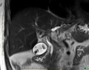
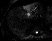
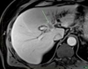
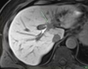


There is marked dilatation of the left hepatic duct, with a subtle, minimally enhancing mass in the porta hepatis. CBD is not dilated. Delayed imaging with Primovist (hepatocyte agent) shows the right lobe hepatocytes enhancing maximally, less marked enhancement in the left lobe due to chronic obstruction and minimally enhancing mass in the hilum consistent, which also shows restricted diffusion.
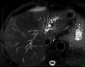
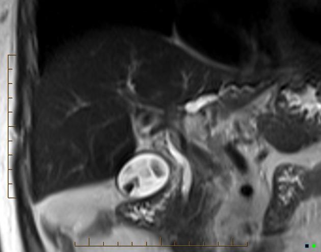
MRCP showing typical stricture at the junction of the right and left hepatic ducts extending into the common hepatic duct (causing the most significant obstruction to the left hepatic duct).
Interpretation: Imaging features are highly likely representing cholangiocarcinoma (Klatskin tumor).
Case Discussion
Features highly suggestive for a hilar cholangiocarcinoma, which is also referred as Klatskin tumor.


 Unable to process the form. Check for errors and try again.