
Presentation
Biopsy Proven Langerhans cell histiocytosis
Patient Data

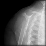
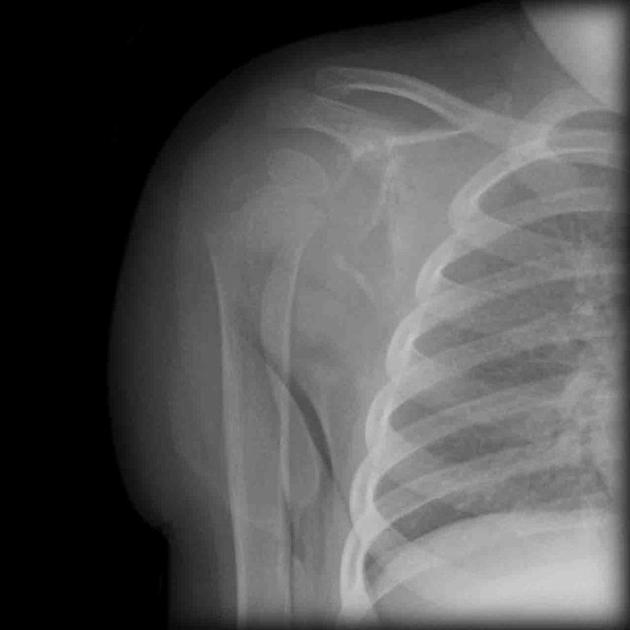
The x-rays both AP and oblique demonstrates osseous destruction in the right shoulder region.

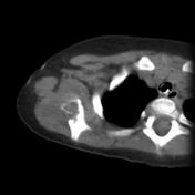
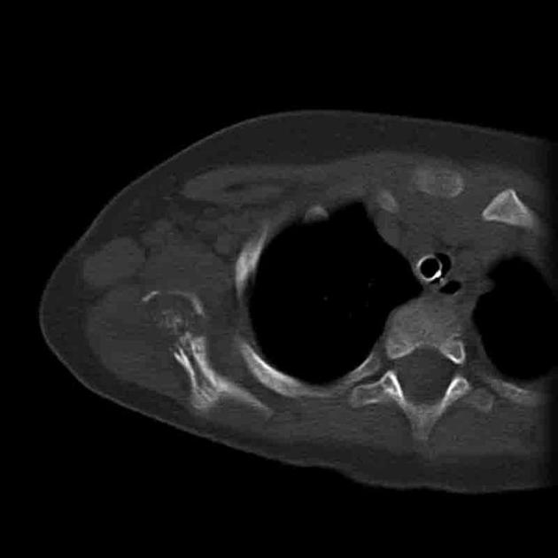
The CT (bone view) and MR images (coronal and axial) are able to better delineate the extent of destruction and any associated surrounding tissue involvement.

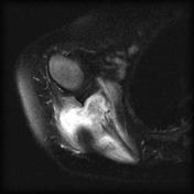
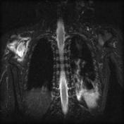
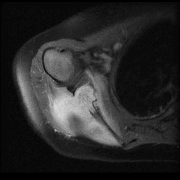
The CT (bone view) and MR images (coronal and axial) are able to better delineate the extent of destruction and any associated surrounding tissue involvement.
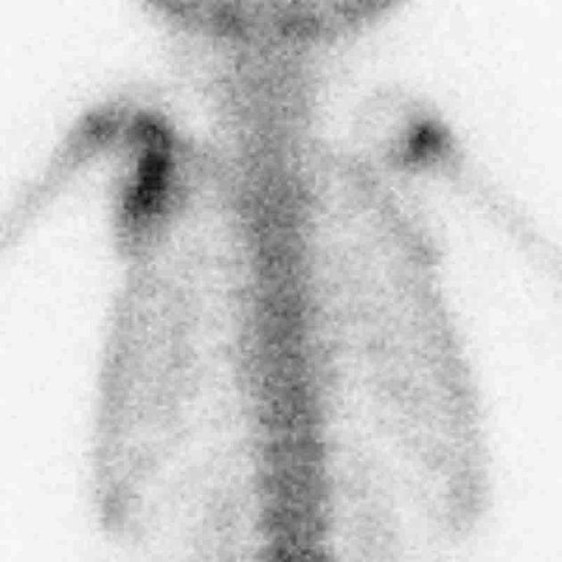
Nuclear medicine scan demonstrates increasing uptake in the right shoulder region.
Case Discussion
This is a case of biopsy proven Langerhans Cell Histiocytosis in an 18 month old child.
Langerhans cell histiocytosis is a condition where excess immune cells (Langerhans cells) accumulate. These cells are important for the appropriate functioning of the immune systems and are found in the spleen, lymph nodes and bone marrow. This condition can be biopsy proven.
Images provided by: Dr Frank Gaillard


 Unable to process the form. Check for errors and try again.