
Presentation
Pelvic mass.
Patient Data
Age: 50 years
Gender: Female
From the case:
Large ovarian fibroma


Download
Info

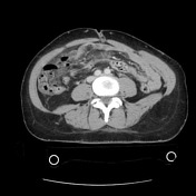
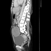
Large well-circumscribed homogeneous central pelvic mass starting below the iliac bifurcation, indenting the bladder dome and anterior body wall.
Case Discussion
Ultrasound-guided biopsy was safely performed through the body wall with initial pathology suspecting spindle cell neoplasm (possible desmoid tumor). In fact, at the time of surgery, this was attached to the left ovary, which is quite difficult to determine by CT and the final pathologic diagnosis is a benign ovarian fibroma.


 Unable to process the form. Check for errors and try again.