
Presentation
History of preterm birth at 30 weeks of gestation. Hospitalized for 10 days during birth. Presenting with multiple episodes of seizures and global developmental delay.
Patient Data
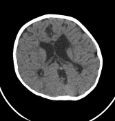

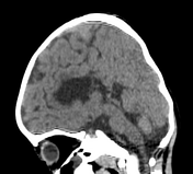

Mild dilatation of bilateral lateral ventricles predominantly frontal horn and body with irregular margin and severe thinning or loss of periventricular and deep white matter; mildly prominent bilateral cortical sulci and cisterns – features of parenchymal volume loss.
Few subcentimeter areas of CSF attenuation in bilateral periventricular region – likely gliosis or periventricular cysts.
Tiny punctate calcification in the right side – likely dystrophic calcification. Thinned out corpus callosum.
Above features likely sequala of hypoxic ischemic encephalopathy.
Case Discussion
CT and MR imaging findings of end-stage PVL include ventriculomegaly with irregular margins of the bodies and trigones of the lateral ventricles, loss of periventricular white matter with increased T2 signal, and thinning of the corpus callosum.


 Unable to process the form. Check for errors and try again.